Development and validation of an intra-tumoral and peri-tumoral radiomics model based on dynamic contrast-enhanced ultrasound for predicting lymph node metastasis in type 2 diabetic patients with thyroid cancer - Summary - MDSpire
Advertisement
Development and validation of an intra-tumoral and peri-tumoral radiomics model based on dynamic contrast-enhanced ultrasound for predicting lymph node metastasis in type 2 diabetic patients with thyroid cancer
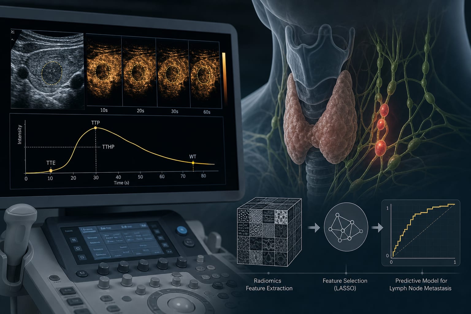
To develop and validate intra-tumoral and peri-tumoral radiomics models based on dynamic contrast-enhanced ultrasound (CEUS) to preoperatively predict lymph node metastasis (LNM) in thyroid cancer patients with type 2 diabetes.
Approach:
Key Findings:
The 2 mm peri-tumoral region yielded a higher AUC than the 1 mm region in all cohorts, with statistical significance in the training cohort and a consistent trend in the external validation cohorts.
The combined radiomics model achieved AUCs of 0.930 (95% CI: 0.876–0.964), 0.907 (95% CI: 0.796–0.968), and 0.865 (95% CI: 0.739–0.941) in the training and external validation cohorts.
Calibration curves showed good agreement between predicted and actual outcomes.
The CEUS-based combined radiomics model using intra-tumoral and 2 mm peri-tumoral features provides an effective tool for preoperative LNM prediction in thyroid cancer patients with type 2 diabetes.
Conclusion:
The study presents a non-invasive and accurate preoperative assessment tool for predicting LNM in high-risk diabetic populations.