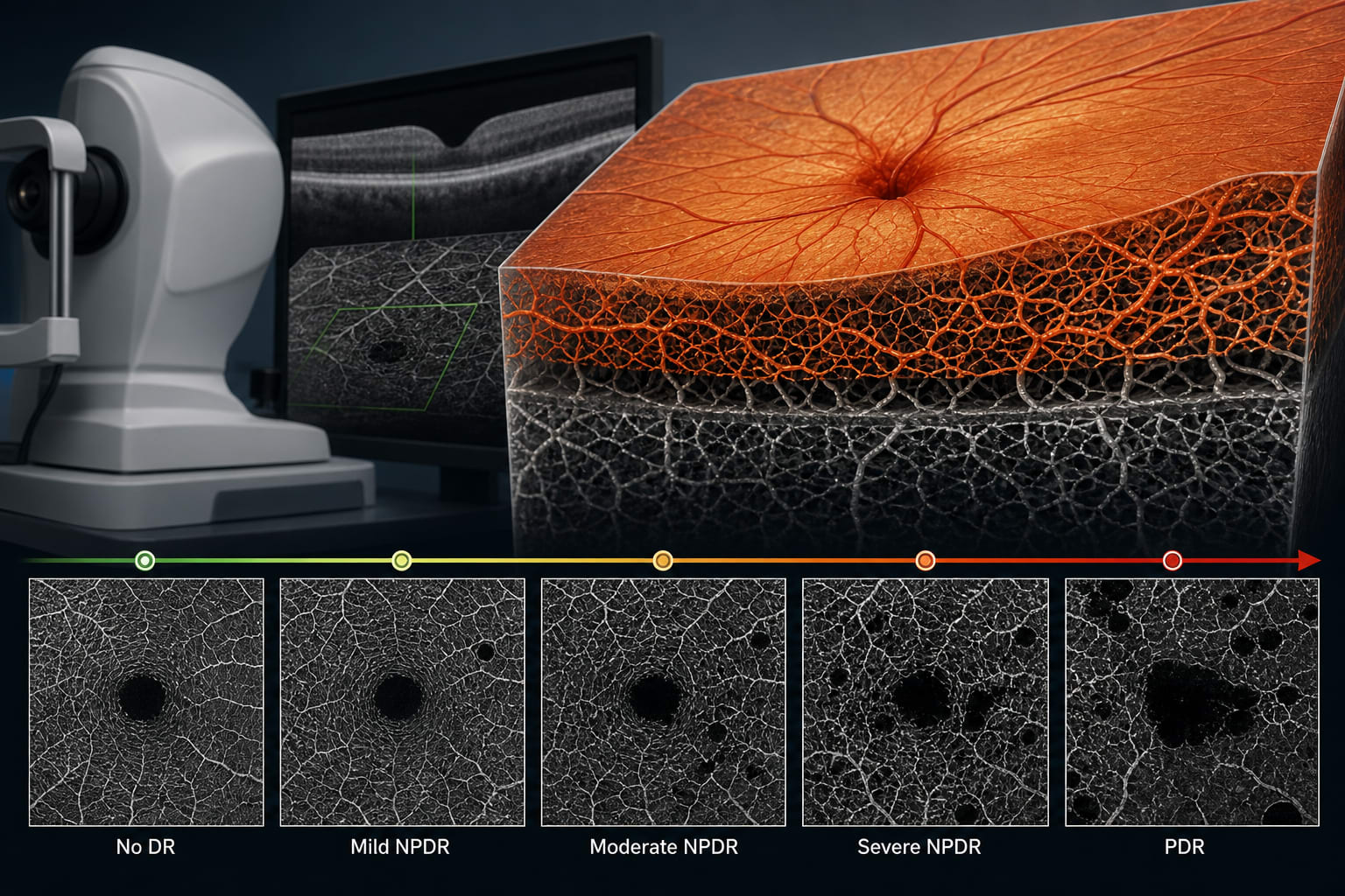
To identify OCTA-derived biomarkers correlating with diabetic retinopathy (DR) severity and characterize longitudinal retinal microvascular changes across DR stages.
Approach:
Study Design: A 3-year prospective study analyzing OCTA images from 328 eyes of 164 adults with type II diabetes and 33 eyes from 17 healthy controls.
Data Collection: Bilateral OCTA scans were obtained at each visit, and seven microvascular indices were extracted from the superficial and deep capillary plexuses.
Statistical Analysis: Linear regression quantified changes over time, with intergroup comparisons made using ANOVA and post-hoc testing.
Key Findings:
In the deep capillary plexus, eyes with severe NPDR exhibited greater annual declines in vessel density (-0.563 ± 0.39% per year) and skeleton density (-0.296 ± 0.23% per year) compared with controls.
Acircularity index increased over time, with greater annual increases observed in severe NPDR relative to mild NPDR (0.16 ± 0.12% per year), consistent with progressive macular ischemia.
In the superficial plexus, eyes with severe NPDR showed greater annual reductions in average vessel caliber (-4.5e-4 ± 0.0005% per year) compared with earlier disease stages.
Most OCTA metrics demonstrated small but statistically significant longitudinal trends.
Interpretation:
Longitudinal OCTA analysis reveals stage-specific microvascular changes in DR.
Limitations:
The study's design did not allow for re-grading of DR severity during follow-up.
Current longitudinal studies on OCTA and DR are limited in breadth and scope, often including only one stage of DR or only one microvasculature metric.
Conclusion:
OCTA-derived biomarkers provide a noninvasive tool for detecting early DR changes.
An artificial intelligence–based optical coherence tomography pathway met noninferiority criteria for false-positive diabetic macular edema referrals and was associated with fewer referral decisions in a randomized clinical trial.