To describe two patients with numerous, localized basal cell carcinomas (BCCs) without clinical features of an associated genetic syndrome, and discuss diagnostic and treatment challenges.
Approach:
Case 1: A 67-year-old woman developed 22 BCCs over 24 years, including 19 on the frontal scalp. She had a posterior-neck port wine stain and denied unusual sun exposure or sunburns, immunosuppression, family history of skin cancer, and radiation, trauma, injury, or chemical contact involving the affected areas. No associated syndromic stigmata were identified.
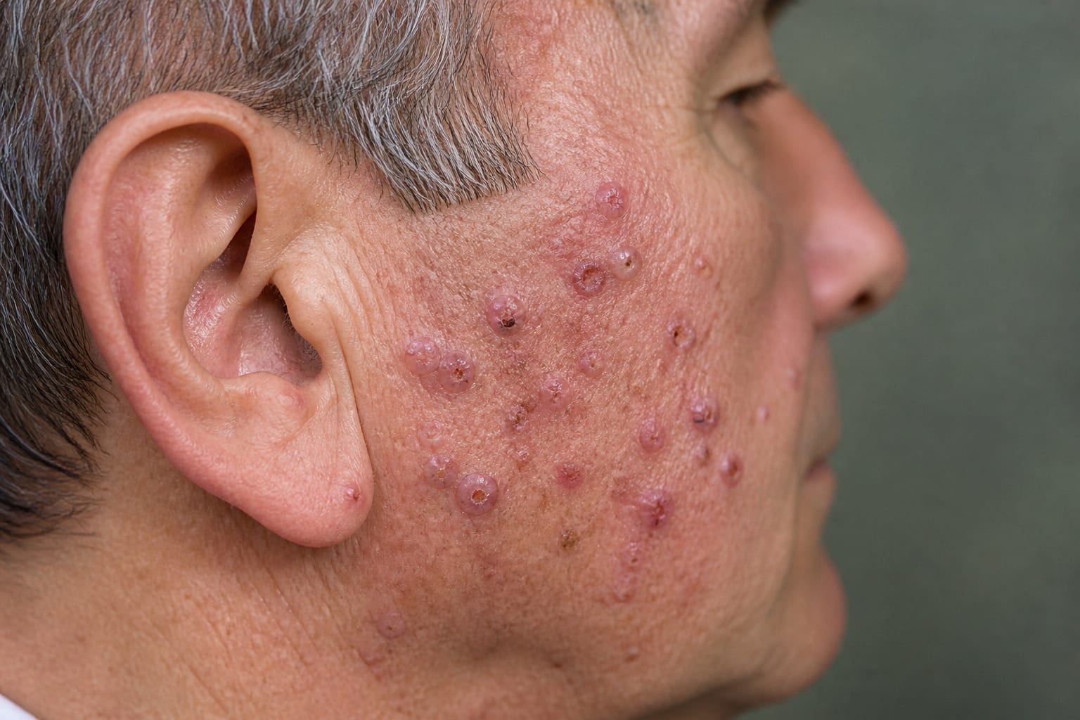
Case 2: A 78-year-old man developed 44 BCCs over 11 years. Twenty-four involved the right side of the face, including 20 clustered in the right perioral and buccal region. He had received radiation to the entire face for acne as an adolescent. No associated syndromic stigmata were identified.
Diagnosis and management: External dermatopathologists diagnosed each lesion, and the diagnoses were confirmed during Mohs micrographic surgery. Both patients received combinations of electrodesiccation and curettage, Mohs micrographic surgery, and standard excision.
Key Findings:
Both patients had numerous histologically confirmed BCCs concentrated in a localized region and lacked identified clinical stigmata of BCC-associated genetic syndromes.
In both cases, lesions accumulated progressively over more than a decade.
The authors discuss localized cutaneous mosaicism as a possible explanation; sequencing of affected skin was not performed, so the hypothesis remains unconfirmed.
The second patient’s predominantly right-sided facial tumors were not fully accounted for by his history of radiation to the entire face.
Interpretation:
The authors describe both cases as nonsyndromic agminated BCC. Because the lesions developed progressively over more than a decade, the cases raise the question of whether a short time interval should be part of the definition of agminated BCC.
Limitations:
DNA from affected skin was not sequenced, so cutaneous mosaicism could not be assessed.
The report describes only two patients.
Conclusion:
The cases illustrate diagnostic and treatment challenges when multiple BCCs develop in a localized area without an apparent syndromic context.
Immunopeptidomics is helping researchers identify tumor-presented antigens that could inform the development of targeted cell therapies and personalized cancer vaccines