To establish a quantitative framework based on MRI data for stratifying the complexity of glioma imaging.
Approach:
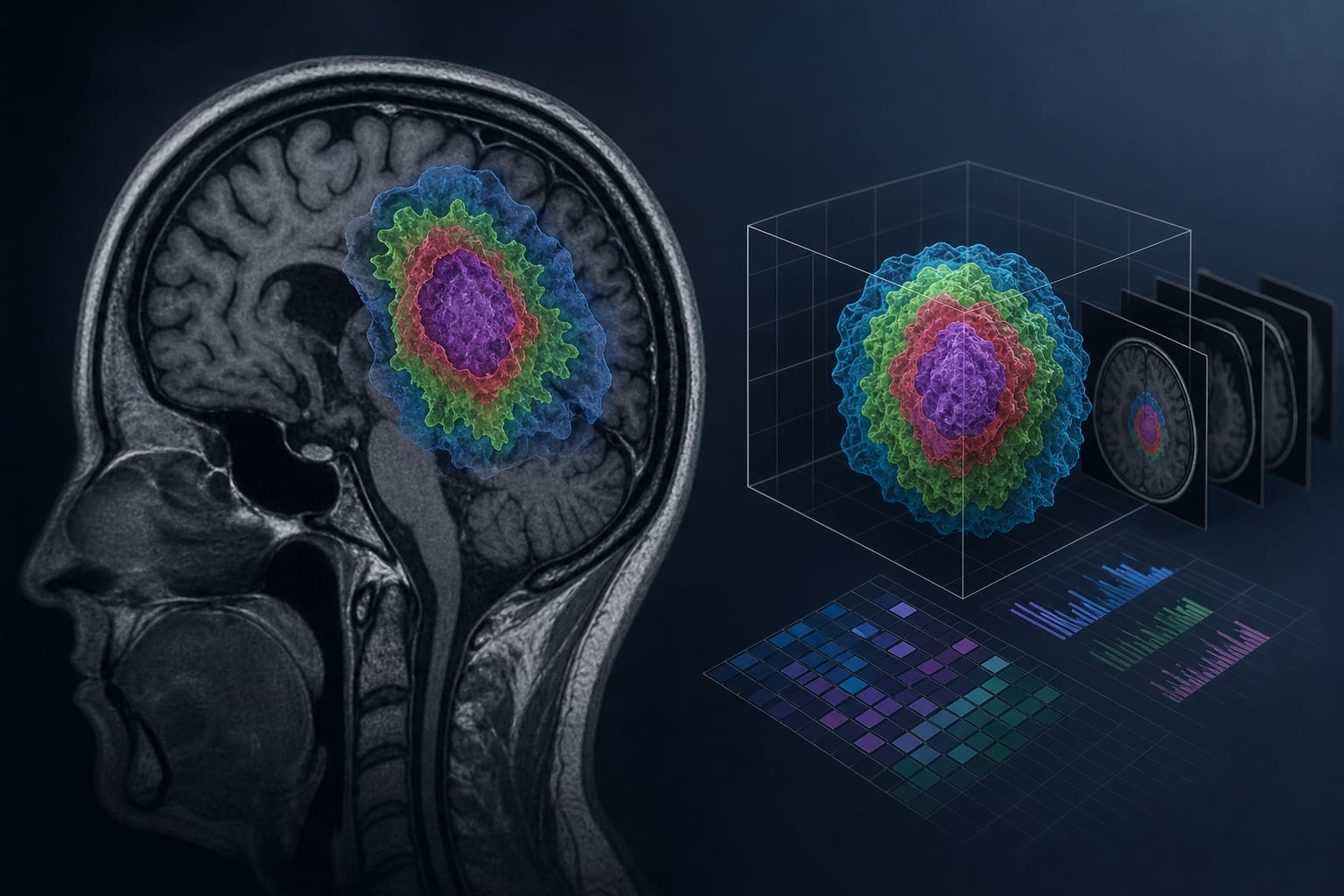
Retrospective Analysis: A retrospective quantitative MRI analysis was performed using segmentation datasets from 1,251 glioma cases obtained from the BraTS repository.
Quantitative Imaging Variables: Quantitative imaging variables were extracted using voxel-based segmentation methods and included total tumor volume, enhancing tumor volume, edema volume, necrotic/non-enhancing core volume, and maximum tumor diameter.
Statistical Analysis: Statistical analysis included ANOVA, Kruskal–Wallis testing, and Pearson/Spearman correlation analysis.
Key Findings:
Three evenly distributed groups were created, each comprising 417 cases.
Mean total tumor volume increased across GICS categories from 35.40 ± 15.84 cm3 in GICS-1 to 162.62 ± 34.96 cm3 in GICS-3 (p < 0.001).
Higher GICS categories correlated with greater edema burden, increased maximum tumor diameter, and larger enhancing and necrotic components.
Strong positive correlations were found between total tumor volume and maximum tumor diameter (Pearson r = 0.764; p < 0.001) and between total tumor volume and edema volume (Pearson r = 0.861; p < 0.001).
Interpretation:
The GICS framework represents a preliminary quantitative MRI stratification model using standardized segmentation datasets, indicating that segmentation-derived MRI variables can be organized into reproducible stratification groups.
Limitations:
The study is based on retrospective data.
The framework requires further validation in prospective studies.
Conclusion:
The GICS framework lays the groundwork for prospective studies on imaging-based complexity assessment and exploratory visualization-support approaches in glioma surgery.