AI-Assisted MRI Method May Rival CT in Lung Nodule Detection
A rapid AI‐assisted imaging protocol detected nearly all clinically relevant solid lung nodules while closely matching CT for suspicious screening classifications.
-
By
-
Andrea Surnit
-
August 5, 2026
-
Objective:
To evaluate the diagnostic performance of an AI-assisted MRI protocol for detecting pulmonary nodules compared to CT.
Approach:
- Study Design: A single-center prospective study involving 97 asymptomatic adult patients who underwent whole-body MRI and same-day CT.
- Imaging Protocol: Utilized a single-breath-hold, AI-assisted compressed-sensing three-dimensional T1-weighted fast field echo sequence.
Key Findings:
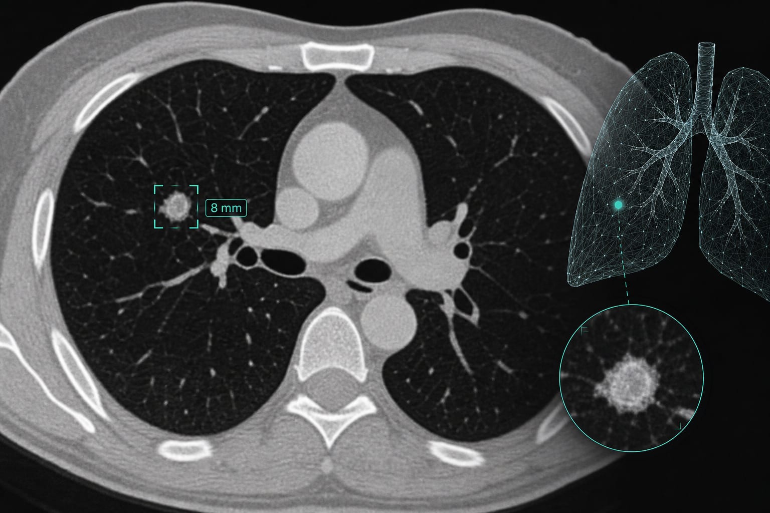
- CT identified 118 pulmonary nodules, while MRI detected 78 with one false-positive.
- MRI achieved 83% sensitivity and 87% accuracy for solid and part-solid nodules.
- For solid nodules ≥4 mm, MRI sensitivity and accuracy reached 98%.
- No clinically relevant suspicious nodules were missed in the cohort.
- MRI showed reduced sensitivity for nodules <4 mm and calcified nodules.
Interpretation:
Limitations:
- Conducted at a single center with a small cohort, limiting generalizability.
- Reduced sensitivity for nodules <4 mm, particularly calcified nodules.
- Requires validation in larger screening cohorts and across multiple imaging platforms.
Conclusion:
Sources: