To characterize choroidal structural and perfusion changes in children with emmetropia, low myopia, and moderate myopia using enhanced depth imaging optical coherence tomography (EDI-OCT) and optical coherence tomography angiography (OCTA), and to examine their associations with spherical equivalent refraction (SER) and axial length (AL).
Key Findings:
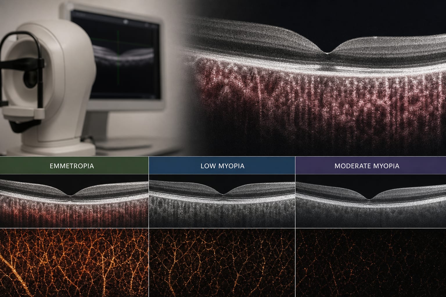
Choroidal thickness (CT) decreased significantly with increasing myopia severity at all measured macular locations.
The reduction in CT was primarily due to thinning of Haller’s layer.
Significant reductions in luminal area (LA) and total choroidal area (TCA) were observed.
No significant differences in OCTA-derived perfusion or flow-deficit parameters between groups.
OCT-derived structural parameters were positively correlated with SER and negatively correlated with AL.
Interpretation:
Limitations:
The study was limited to a single center and a specific age range.
Only right-eye data were included to avoid inter-eye correlation.
Conclusion:
Structural OCT-based metrics may better reflect early choroidal changes across refractive groups in children than OCTA-derived perfusion parameters.