To illustrate a case of appendiceal abscess presenting as an isolated abdominal wall mass without abdominal pain, and to highlight the diagnostic challenges encountered.
Approach:
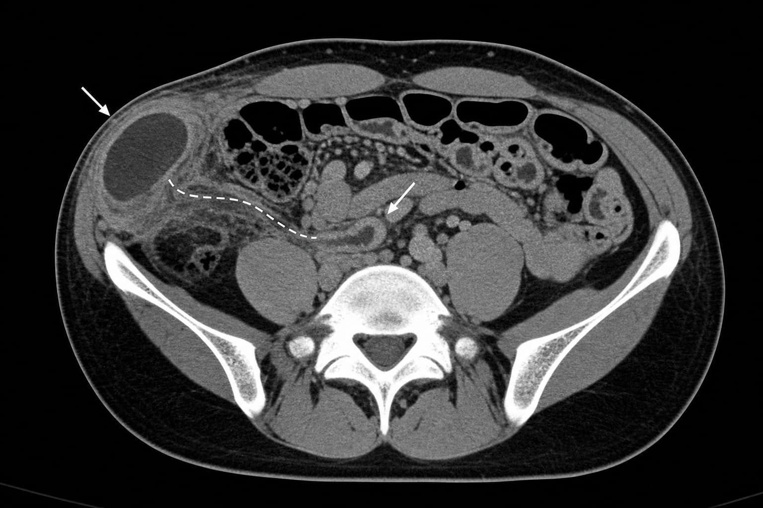
Case Presentation: A 13-year-old boy presented with a 10-day history of a painful right lower abdominal wall swelling, explicitly denying any abdominal pain. Initially misdiagnosed with a soft tissue infection based on a focused abdominal wall ultrasound, he was referred after 10 days of unsuccessful treatment. A non-contrast computed tomography scan revealed a secondary abdominal wall abscess connected via an inflammatory tract to a large periappendiceal abscess.
Key Findings:
An isolated abdominal wall mass in a child with systemic inflammation may indicate an intra-abdominal source, even without abdominal pain.
Comprehensive abdominal ultrasound is essential to avoid missing occult intra-abdominal pathology.
In cases of delayed presentation, cross-sectional imaging (CT or MRI) is warranted if ultrasound results are inconclusive.
Staged management with antibiotics followed by interval appendectomy is effective for complex appendicitis.
Interpretation:
This case illustrates a diagnostic pitfall where the absence of expected abdominal pain led to misdiagnosis, highlighting the need for thorough evaluation in cases of unexplained abdominal wall masses.
The case serves as a reminder for clinicians to maintain a broad differential diagnosis and adhere to imaging guidelines in atypical presentations of appendicitis.
The procedure was performed under a HOPE Act research protocol at an NYU Langone Health center the institution said is among the limited number of US transplant centers equipped and approved to perform HOPE lung transplants.