To investigate the clinical and dermoscopic features of acquired facial hyperpigmented macules (AFHM) and their significance in pediatric dermatology.
Approach:
Key Findings:
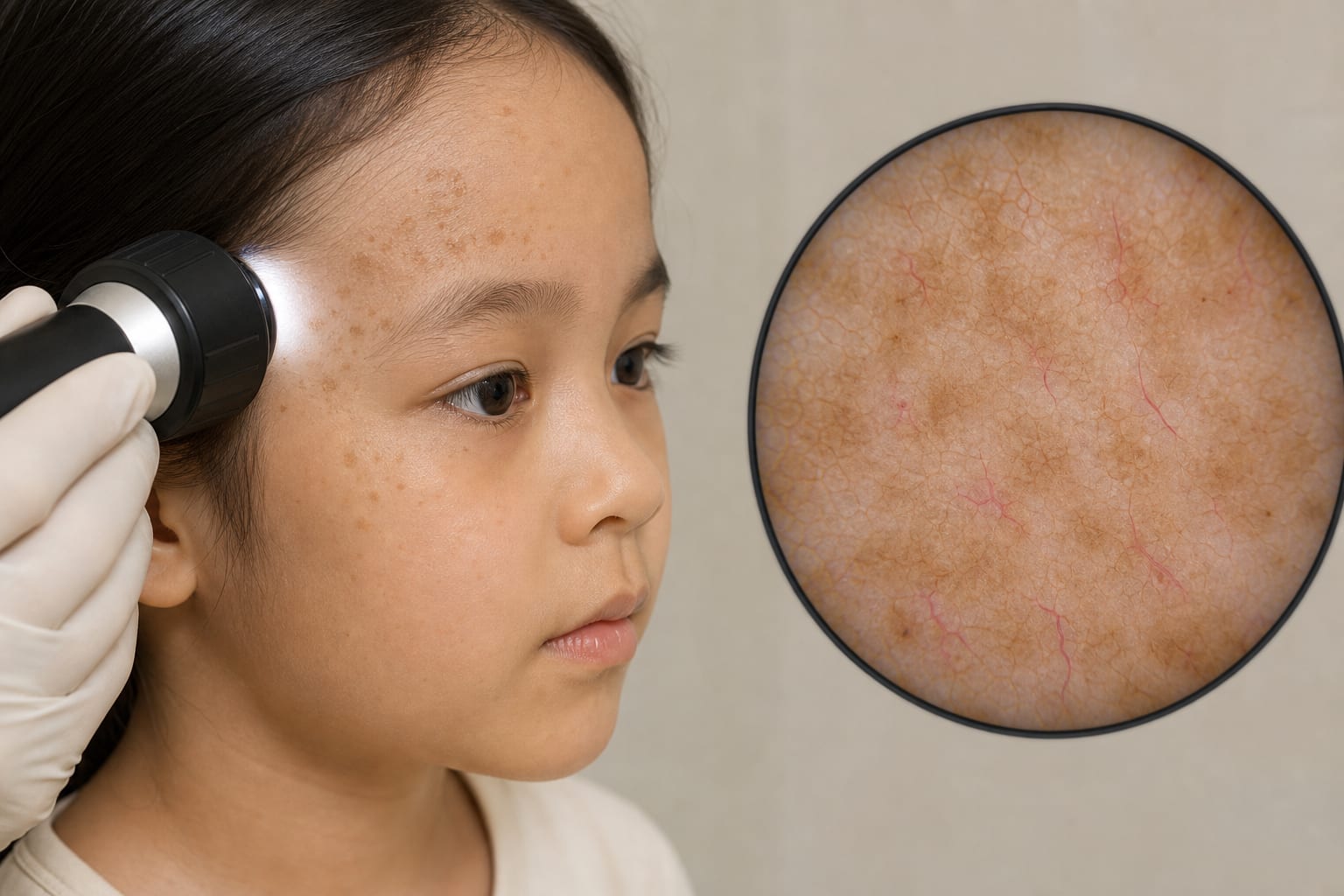
Lesions are light brown macules, irregularly shaped, ranging from millet to peanut size, primarily located on the forehead and temples.
Major dermoscopic features include light brown pseudoreticular pigment and linear/branching vessels.
During follow-up, 96.4% of 28 patients showed complete disappearance of lesions.
Interpretation:
AFHM is characterized as a self-limited condition with a potential role of ultraviolet radiation in its etiology, highlighting the need for awareness in pediatric care.
Limitations:
The study is retrospective and may have selection bias.
Limited sample size may affect the generalizability of findings.
Potential biases inherent in retrospective studies should be acknowledged.
Conclusion:
AFHM is a self-limited disease with a high rate of spontaneous resolution in young children, emphasizing the importance of monitoring and sun protection.