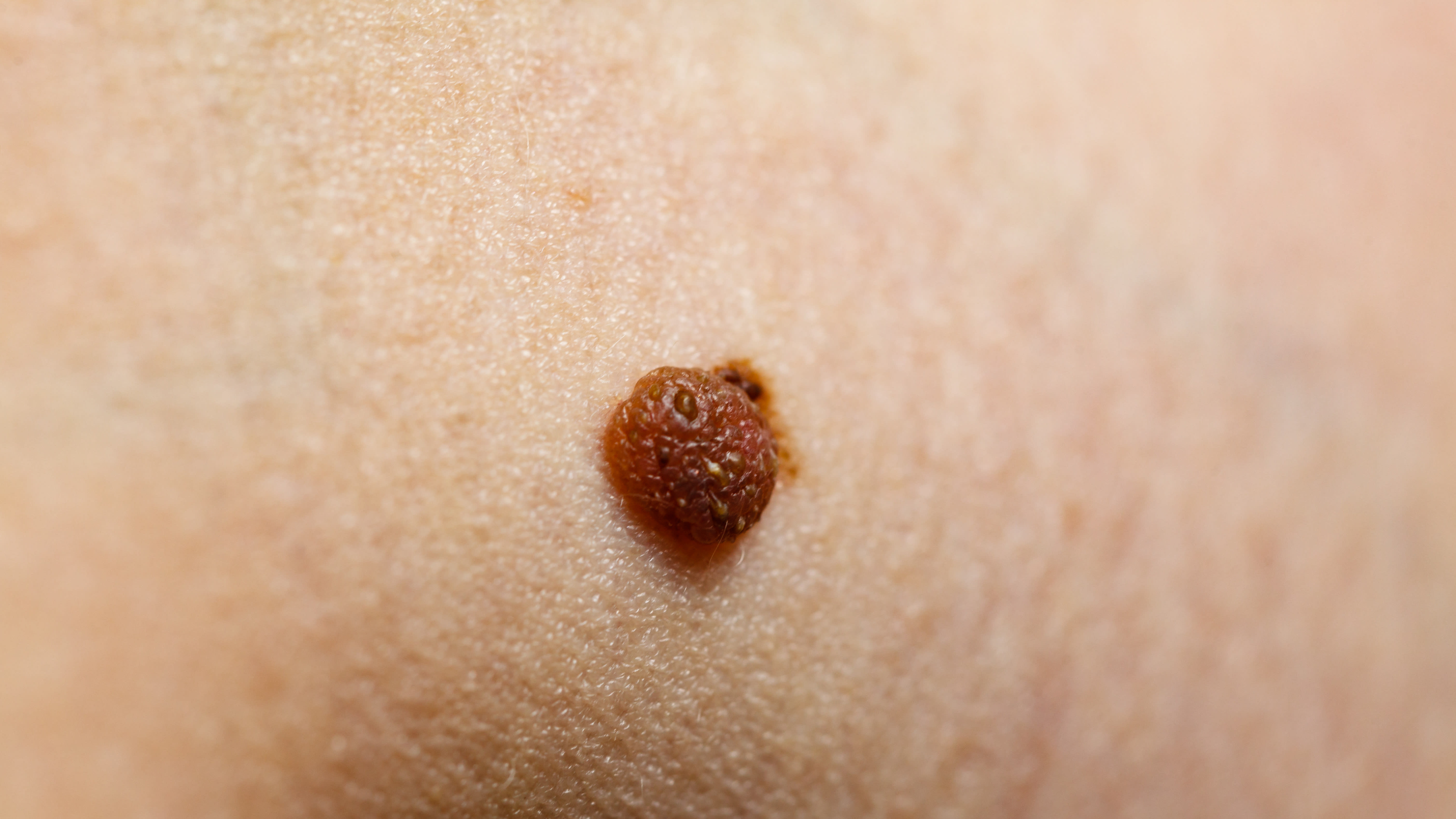
High-frequency ultrasound combined with superb microvascular imaging for differential diagnosis of basal cell carcinoma and seborrheic keratosis: a retrospective analysis with nomogram implementation - Summary - MDSpire
Advertisement
High-frequency ultrasound combined with superb microvascular imaging for differential diagnosis of basal cell carcinoma and seborrheic keratosis: a retrospective analysis with nomogram implementation
To evaluate the diagnostic performance of high-frequency ultrasound (HFUS) combined with superb microvascular imaging (SMI) in differentiating basal cell carcinoma (BCC) from seborrheic keratosis (SK), and to develop a nomogram for individualized probability estimation, emphasizing its non-invasive nature.
Key Findings:
Significant differences between BCC and SK in age, lesion location, size, depth, morphology, internal echotexture, posterior acoustic features, and microvascular flow patterns (all p < 0.001).
Independent predictors of BCC included larger tumor size, head location, nodular ultrasound pattern, and random vascular flow.
The multivariate logistic regression model demonstrated an AUC of 0.861, with sensitivity of 77.55% and specificity of 82.38%.
Interpretation:
HFUS combined with SMI provides a valuable non-invasive approach for differentiating BCC from SK, potentially reducing unnecessary biopsies and facilitating earlier detection of malignant lesions, thereby improving patient outcomes.
Limitations:
Retrospective design may introduce selection bias, potentially affecting the study's conclusions.
Limited to a single institution, which may affect generalizability.
Potential for incomplete data affecting analysis.
Conclusion:
The developed nomogram based on key ultrasound and clinical predictors demonstrates good discriminative performance and offers a practical tool to support clinical decision-making, with implications for future research.