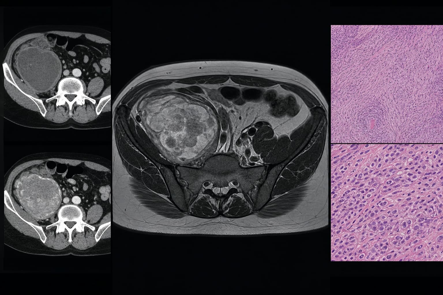
To present a case of primary sclerosing epithelioid fibrosarcoma (SEF) in the iliac fossa, emphasizing its imaging characteristics, diagnostic challenges, and significance in the context of existing literature.
Key Findings:
The case represents the second reported instance of primary SEF in the iliac fossa, with detailed imaging data that highlight its unique characteristics.
Imaging showed a large, heterogeneous mass with specific characteristics that helped differentiate SEF from other tumors, emphasizing the importance of imaging in diagnosis.
No evidence of recurrence or metastasis was observed during a 10-month follow-up, underscoring the need for long-term monitoring.
Interpretation:
The presence of a deep-seated soft-tissue mass with heterogeneous T2 signal and progressive enhancement should prompt consideration of SEF, particularly in atypical locations, highlighting the critical role of imaging in diagnosis.
Limitations:
The follow-up period was short, limiting long-term outcome assessment and potential biases in case reporting.
The rarity of SEF may restrict generalizability of findings.
Conclusion:
Definitive diagnosis of SEF requires histopathology and immunohistochemistry, with long-term follow-up necessary to monitor for recurrence or metastasis, emphasizing the importance of imaging in the diagnostic process.