Case Report: A case of a giant right ventricular wall hematoma caused by coronary artery perforation during percutaneous coronary intervention - Summary - MDSpire
Advertisement
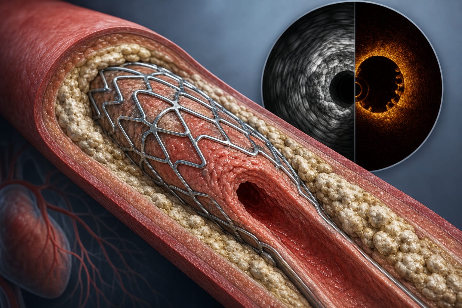
Case Report: A case of a giant right ventricular wall hematoma caused by coronary artery perforation during percutaneous coronary intervention