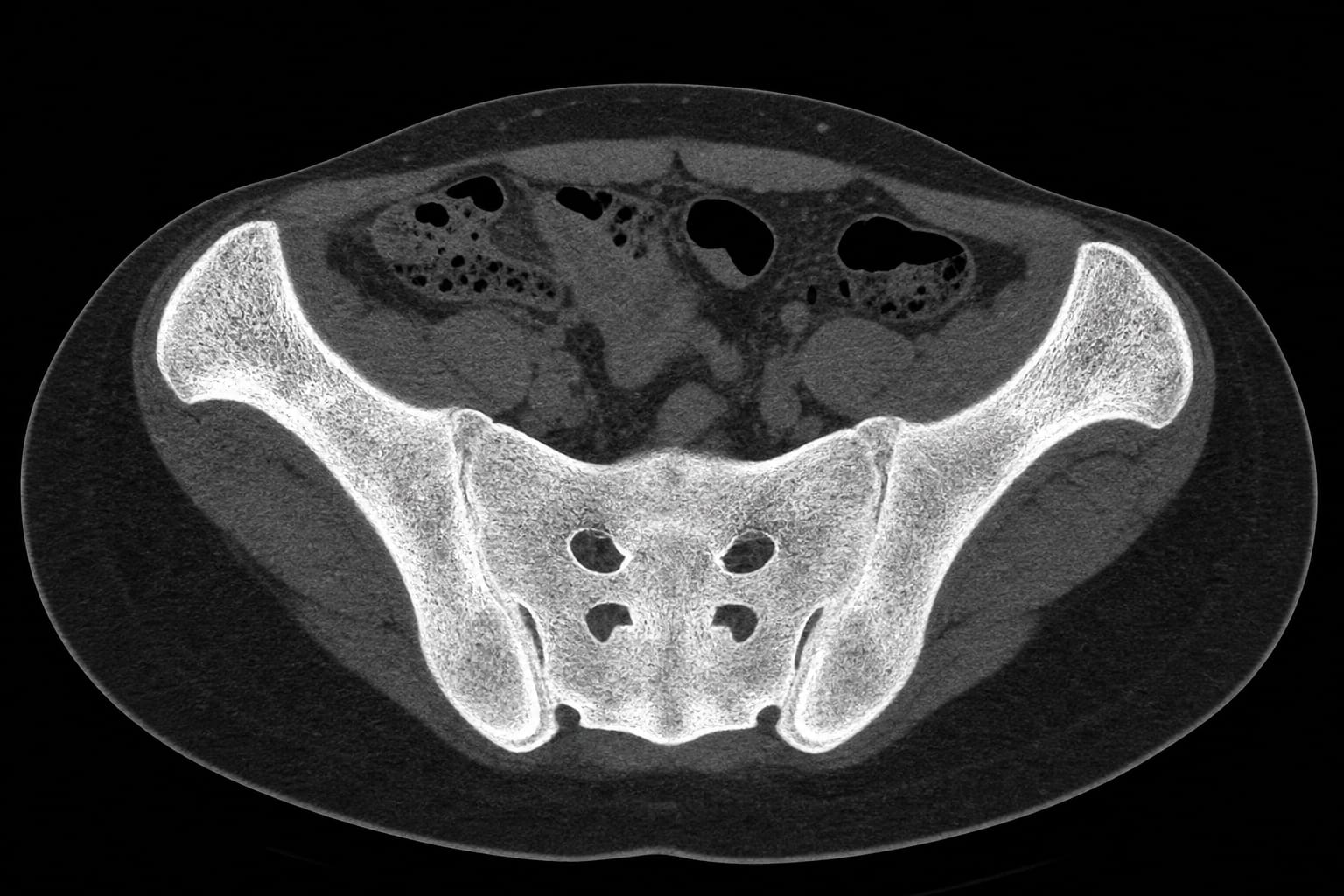
Renal osteodystrophy with near-complete bilateral sacroiliac ankylosis: a bat-like pelvic configuration on CT
-
By
-
Ramazan Orkun Onder
-
Serdar Aslan
-
July 3, 2026
-
Objective:
To present a case of renal osteodystrophy with significant skeletal manifestations, particularly focusing on the CT imaging findings.
Approach:
- Patient Presentation: A 45-year-old woman with chronic kidney disease stage 5D presented with abdominal pain and had a history of hemodialysis.
- Imaging Findings: CT showed bilateral renal atrophy, nephrolithiasis, and diffuse iliac and sacral osteosclerosis with near-complete bilateral sacroiliac ankylosis.
- Biochemical Evaluation: Biochemical tests indicated severe secondary hyperparathyroidism and vitamin D deficiency.
Key Findings:
- CT imaging revealed a bat-shaped pelvic appearance due to bilateral sacroiliac fusion.
- The patient exhibited severe secondary hyperparathyroidism and diffuse axial osteosclerosis.
- Absence of features suggestive of spondyloarthritis and negative HLA-B27 supported the diagnosis of renal osteodystrophy.
Interpretation:
The case illustrates the skeletal manifestations of renal osteodystrophy, particularly the unique CT appearance of sacroiliac ankylosis.
Limitations:
- Previous imaging was unavailable for review, preventing assessment of interval progression.
Conclusion:
This case broadens the recognized CT spectrum of renal osteodystrophy by illustrating near-complete bilateral sacroiliac ankylosis.