To systematically summarize the factors influencing remote diffusion-weighted imaging (R-DWI) lesions after spontaneous intracerebral hemorrhage (ICH) and explore their predictive indicators, prognostic associations, and potential mechanisms.
Approach:
Literature Review: Two reviewers independently searched PubMed, Embase, and the Cochrane Library for relevant studies published in English using specific search terms related to cerebral hemorrhage and diffusion-weighted imaging.
Key Findings:
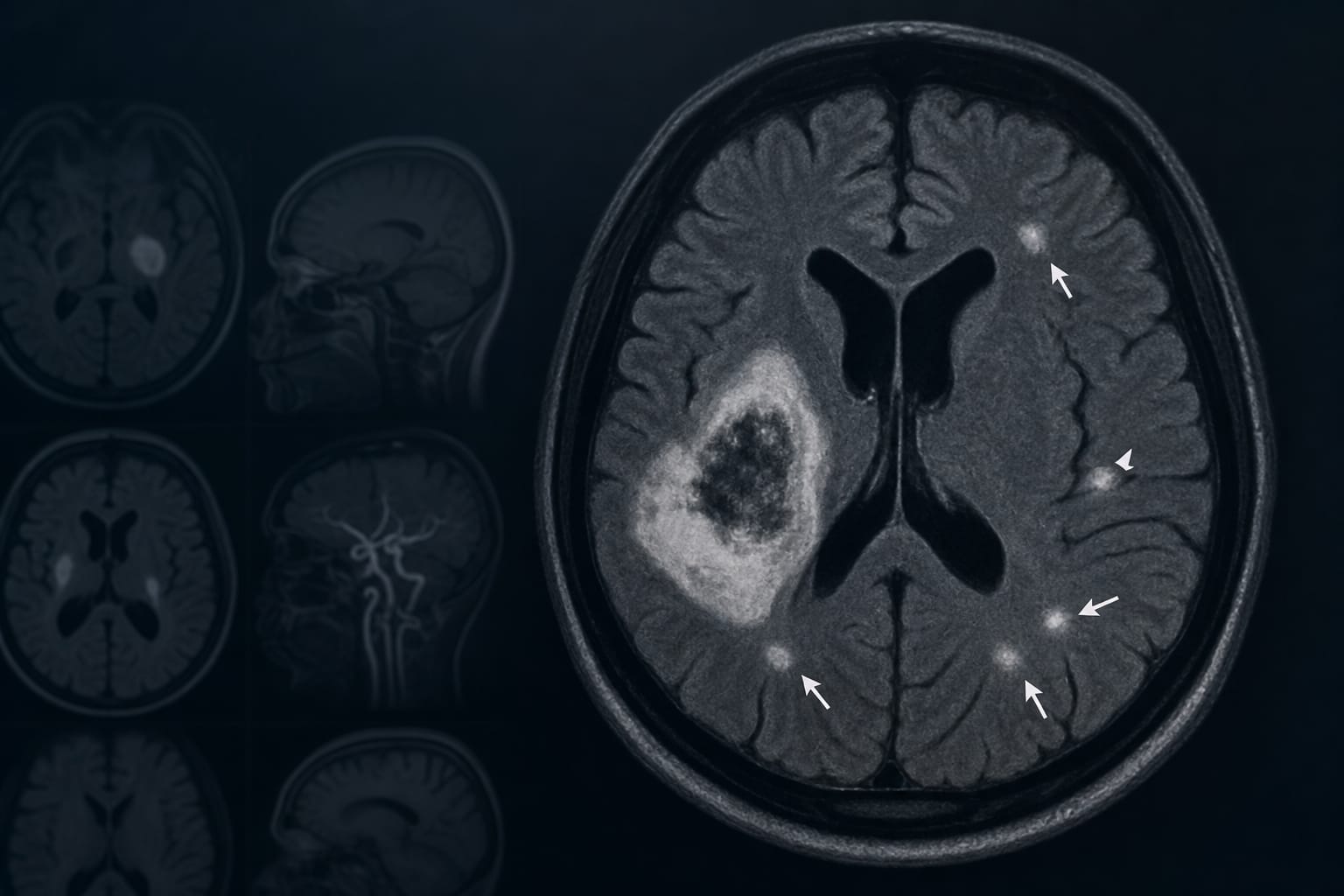
R-DWI lesions are observed in approximately 11-50% of ICH patients.
Patients with R-DWI lesions are often classified into three groups based on underlying conditions: hypertensive arteriopathy, cerebral amyloid angiopathy (CAA), or both.
The pathogenesis of R-DWI lesions is heterogeneous and may involve simultaneous vessel rupture and occlusion, aggressive blood pressure lowering, and prothrombotic/inflammatory processes.
R-DWI lesions are closely linked to the severity of pre-existing cerebral small vessel disease (CSVD), with higher burdens of CSVD markers observed in affected patients.
Interpretation:
The presence of R-DWI lesions may reflect underlying microvascular pathology rather than solely the acute effects of ICH, indicating a need for further research into their mechanisms and implications for patient outcomes.
Limitations:
Current literature on R-DWI lesions is limited and findings are often contradictory.
The mechanisms underlying R-DWI lesions remain unclear and require further investigation.
Conclusion:
Understanding the factors influencing R-DWI lesions could help in developing strategies to improve outcomes for patients with ICH.