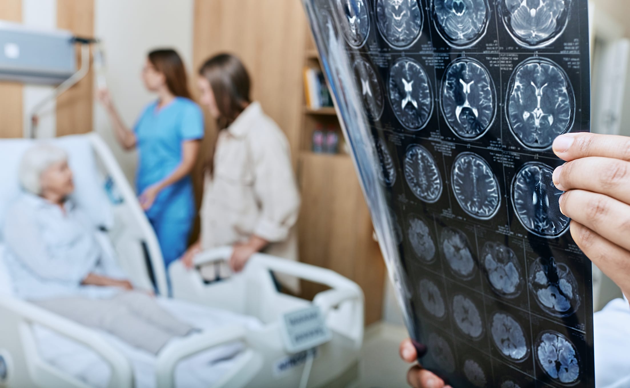
To analyze the relationship between Tmax + 6-second volumes and functional outcomes, specifically focusing on the predictive value of these volumes in patients with mild acute ischemic stroke.
Approach:
Key Findings:
22.9% of patients with mild stroke demonstrated hypoperfusion, indicating substantial tissue at risk, with statistical significance.
Poor functional outcomes at 90 days were significantly associated with Tmax + 6-second volumes of 15 mL or greater.
Functional dependence occurred in 26% of patients with volumes ≥ 15 mL compared to 10% with lower volumes, highlighting the clinical relevance.
Large-vessel occlusion was present in 11.5% of mild stroke patients, emphasizing the need for careful assessment.
Independent predictors of poor outcomes included perfusion lesion volume ≥ 15 mL, hypertension, and large-vessel occlusion, with adjusted odds ratios provided.
Interpretation:
CT perfusion parameters can identify tissue at risk in patients with mild neurological deficits, aiding in early detection of those at risk for poor outcomes.
Limitations:
Retrospective design may introduce selection bias, potentially affecting the generalizability of the findings.
Inclusion limited to patients with available CT perfusion imaging, which may not represent the broader population of mild stroke patients.
Lack of standardization in perfusion thresholds across imaging platforms could lead to variability in results and interpretations.
Conclusion:
CT perfusion can reveal at-risk tissue in mild strokes, facilitating early identification of patients vulnerable to neurological deterioration.