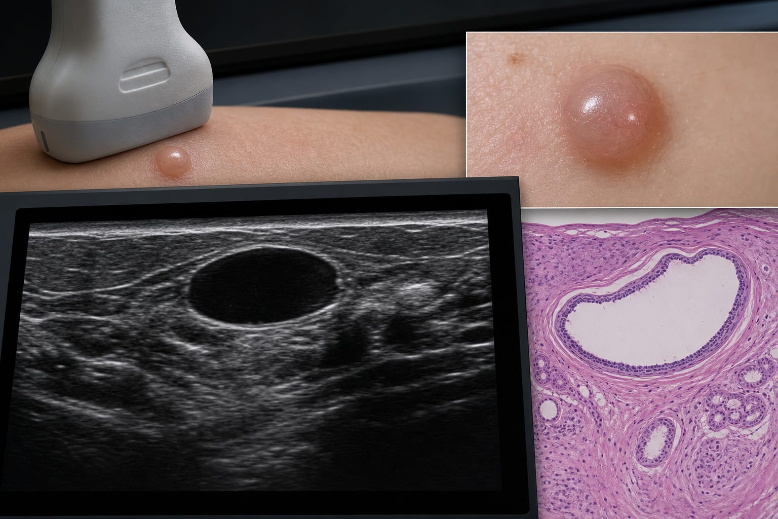
To describe the sonographic features of pathologically confirmed cutaneous hidrocystoma and correlate these findings with histopathologic features.
Approach:
Patient Analysis: Eleven patients with pathologically confirmed cutaneous hidrocystoma were analyzed, undergoing clinical examination and ultrasound evaluation.
Ultrasound Evaluation: Ultrasound examinations were performed by experienced radiologists using high-frequency linear transducers to evaluate various lesion characteristics.
Correlation with Histopathology: Preoperative ultrasound findings were compared with postoperative histopathologic results.
Key Findings:
Eleven lesions were identified in 11 patients, predominantly located in the head and neck region.
Most lesions measured less than 1.5 cm and were located within the dermis or superficial subcutaneous layer.
Sonographic appearances included heterogeneous echogenicity, anechoic cystic features, and hypoechoic characteristics.
Preoperative ultrasound interpretations varied, with none correctly identifying hidrocystoma.
Interpretation:
HFUS features of cutaneous hidrocystoma overlap with atypical epidermoid cysts and hemangiomas.
Limitations:
The study's sample size is small with only 11 patients.
Preoperative ultrasound interpretations did not include hidrocystoma, indicating potential diagnostic challenges.
Conclusion:
HFUS allows precise delineation of lesion depth, internal architecture, and vascular relationships.