Differential infiltration of CD4+ and CD8+ T cells and expression of PD-L1 in paired biopsy and resection specimens of gastric and colorectal adenocarcinomas - Summary - MDSpire
Advertisement
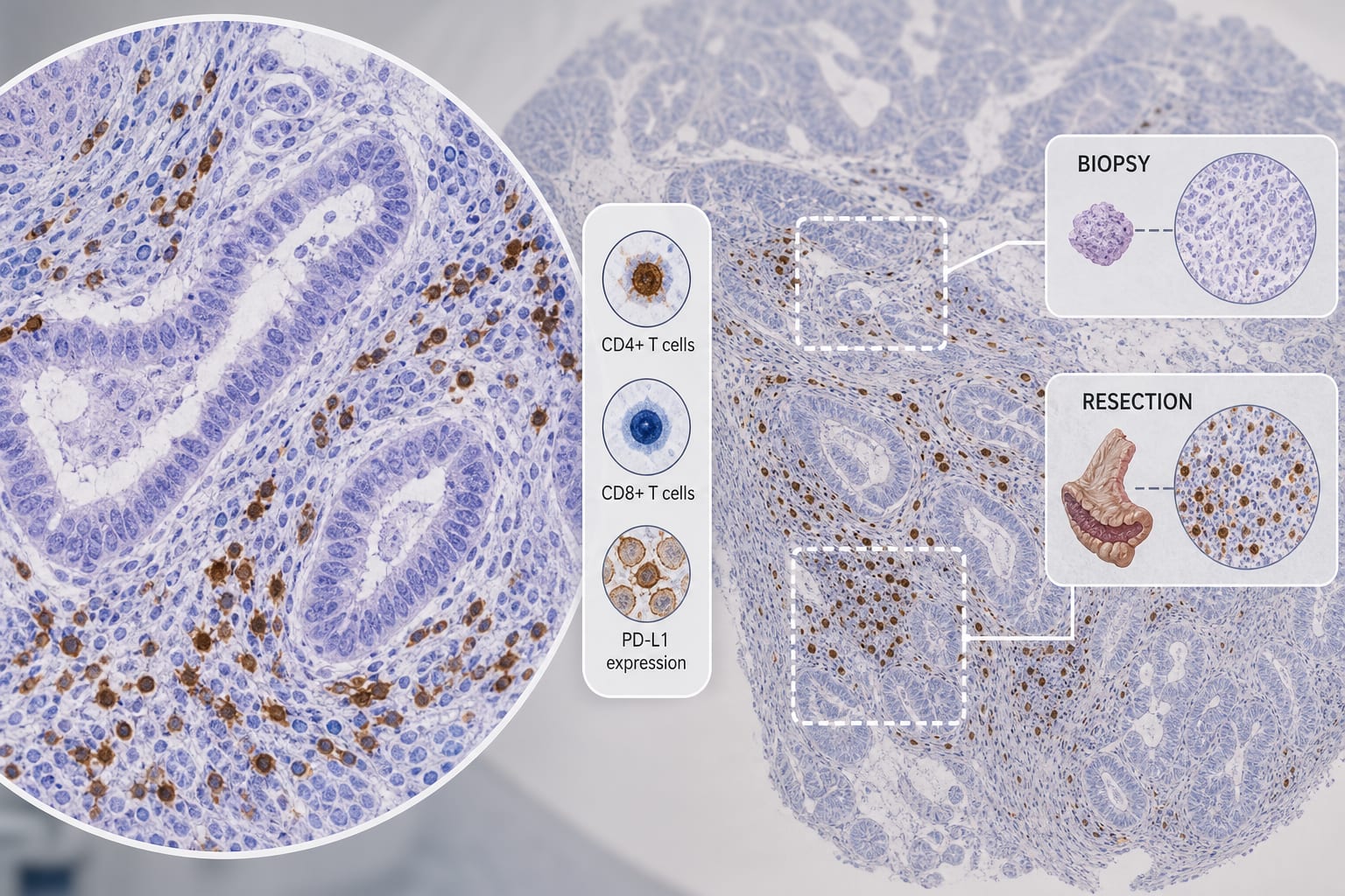
Differential infiltration of CD4+ and CD8+ T cells and expression of PD-L1 in paired biopsy and resection specimens of gastric and colorectal adenocarcinomas
To compare CD4+ T cell, CD8+ T cell infiltration and PD-L1 expression between paired biopsy and resection specimens in gastric and colorectal adenocarcinoma, and evaluate their clinical significance.
Approach:
Key Findings:
In gastric adenocarcinoma, resection specimens showed significantly higher CD4+ T cell, CD8+ T cell density and PD-L1 expression than biopsies (all P<0.05), with CD4+ T cells positively correlated with CA19-9 (R=0.523, P=0.026).
In colorectal adenocarcinoma, only CD8+ T cell density was higher in resection specimens (P=0.0446).
CD4+ T cells negatively correlated with Ki-67 in colorectal adenocarcinoma (R=-0.370, P=0.019).
CD4+/CD8+ ratio negatively correlated with mismatch repair protein expression in colorectal adenocarcinoma (R=-0.342, P=0.029).
Preoperative neutrophil-to-lymphocyte ratio positively correlated with tumor diameter in both cancers.
Interpretation:
Biopsy specimens inadequately represent the immune microenvironment of resection specimens, especially in gastric adenocarcinoma.
Limitations:
ROC analysis demonstrated limited predictive value of biopsy specimens for surgical findings, particularly in colorectal cancer (AUC 0.5-0.7).
Conclusion:
Tumor-type-specific immune assessment of biopsies is essential for guiding precise immunotherapy decisions.