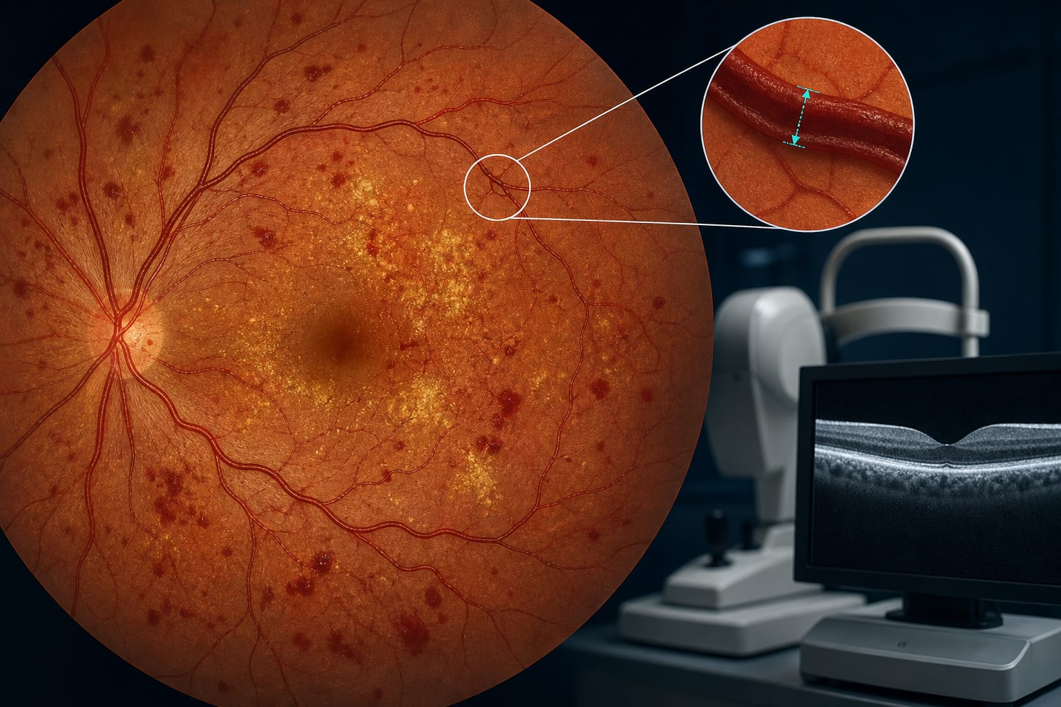
Changes in retinal venous diameter in proliferative diabetic retinopathy patients with diabetic macular edema following faricimab treatment: an observational study - Summary - MDSpire
Advertisement
Changes in retinal venous diameter in proliferative diabetic retinopathy patients with diabetic macular edema following faricimab treatment: an observational study
To evaluate the effect of intravitreal faricimab injections on retinal venous diameter in proliferative diabetic retinopathy (PDR) patients with diabetic macular edema (DME).
Approach:
Study Design: Single-center, retrospective cohort study including 46 PDR patients with DME treated with intravitreal faricimab and panretinal photocoagulation.
Assessment Parameters: Best-corrected visual acuity (BCVA), central foveal thickness (CFT), microaneurysm (MA) count, hard exudates (HE), neovascularization (NV) area, and retinal venous diameter were assessed at baseline and at 1, 3, and 6 months post-treatment.
Key Findings:
Significant improvements in BCVA at 1, 3, and 6 months (P < 0.05).
CFT significantly reduced at all time points (P < 0.05).
MA count showed no significant reduction at 1 month (P > 0.05) but decreased significantly at 3 and 6 months (P < 0.05).
HE area reduction was significant at 6 months (P < 0.05).
NV area significantly reduced at all time points (P < 0.05).
Intravitreal faricimab was safe and effective for PDR patients with DME, showing a trend toward morphological reversal of retinal venous diameter at 6 months.
Limitations:
Retrospective design may introduce bias.
Single-center study limits generalizability.
Small sample size may affect statistical power.
Conclusion:
Faricimab may influence retinal microvascular remodeling through dual inhibition of the VEGF-A and Ang-2 pathways.