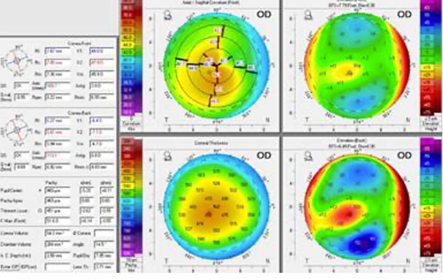
To document the management of keratoconus in a patient through various contact lens fittings over time.
Approach:
Patient History: The patient was first examined in 1997 with toxoplasmosis, amblyopia, and central corneal scarring in the right eye.
Lens Fitting: Initially fitted with a specialized bicurve rigid gas permeable lens for keratoconus, later refitted with large intralimbal GPs in both eyes.
Vision Assessment: Despite the right eye's lack of central vision, the patient reported improved functional peripheral vision.
Lens Specifications: Intralimbal GP lenses were fitted with specific parameters to enhance tear exchange and maintain corneal health.
Follow-Up: In 2019, contact lens wear was deemed contraindicated for the right eye due to corneal integrity risks, leading to a refit with a scleral lens.
Key Findings:
The patient experienced improved peripheral vision despite advanced keratoconus.
Intralimbal GP lenses provided stability and comfort while maintaining corneal health.
Modern scleral lenses offer functional vision improvements even in eyes with poor central acuity.
Interpretation:
This case highlights the patient's experience with different contact lens technologies and their impact on vision.
Limitations:
The study is based on a single patient case, limiting generalizability.
Long-term outcomes of scleral lens use were not detailed.
Conclusion:
The case demonstrates how specific contact lens designs have improved functional vision for a patient with keratoconus.
Recognizing the signs of corneal ectasia can help with prevention of vision loss, and may decrease the emotional impact and functional deficits that can follow.