Intratumoral and peritumoral MRI habitat imaging for differentiating stage IA endometrial cancer from benign endometrial lesions: a multicenter study - Summary - MDSpire
Advertisement
Intratumoral and peritumoral MRI habitat imaging for differentiating stage IA endometrial cancer from benign endometrial lesions: a multicenter study
To evaluate the value of different multiparametric MRI-based radiomics models in differentiating stage IA endometrial cancer (EC) from benign endometrial lesions.
Approach:
Study Design: A retrospective study involving 787 patients with stage IA EC or benign endometrial lesions from four centers.
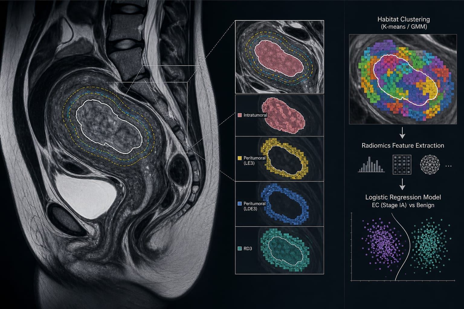
MRI Analysis: Manual delineation of tumor ROIs on MRI, with automatic generation of peritumoral ROIs using Python.
Modeling Techniques: Habitat clustering performed using K-means and Gaussian Mixture Model (GMM) algorithms, with logistic regression to identify predictors.
Performance Evaluation: Diagnostic performance assessed using the area under the curve (AUC).
Key Findings:
Age and vaginal bleeding were identified as independent clinical predictors of stage IA EC.
Habitat-based models outperformed the clinical model in some external validation cohorts.
The Clinical+K-means_RD3 model achieved the highest diagnostic performance with a peak AUC of 0.921.
Interpretation:
Intratumoral and peritumoral habitat imaging based on multiparametric MRI can non-invasively reveal the microstructural characteristics of stage IA EC.
Limitations:
The study is retrospective and may have inherent biases.
Results may not be generalizable to all populations due to specific inclusion criteria.
Conclusion:
Integrating clinical predictors with habitat models showed good diagnostic performance, with the peritumoral habitat model exceeding the intratumoral habitat model.