To address the significant challenge ophthalmologists face in identifying, localizing, and assessing peripheral retinal lesions within a single imaging system.
Key Findings:
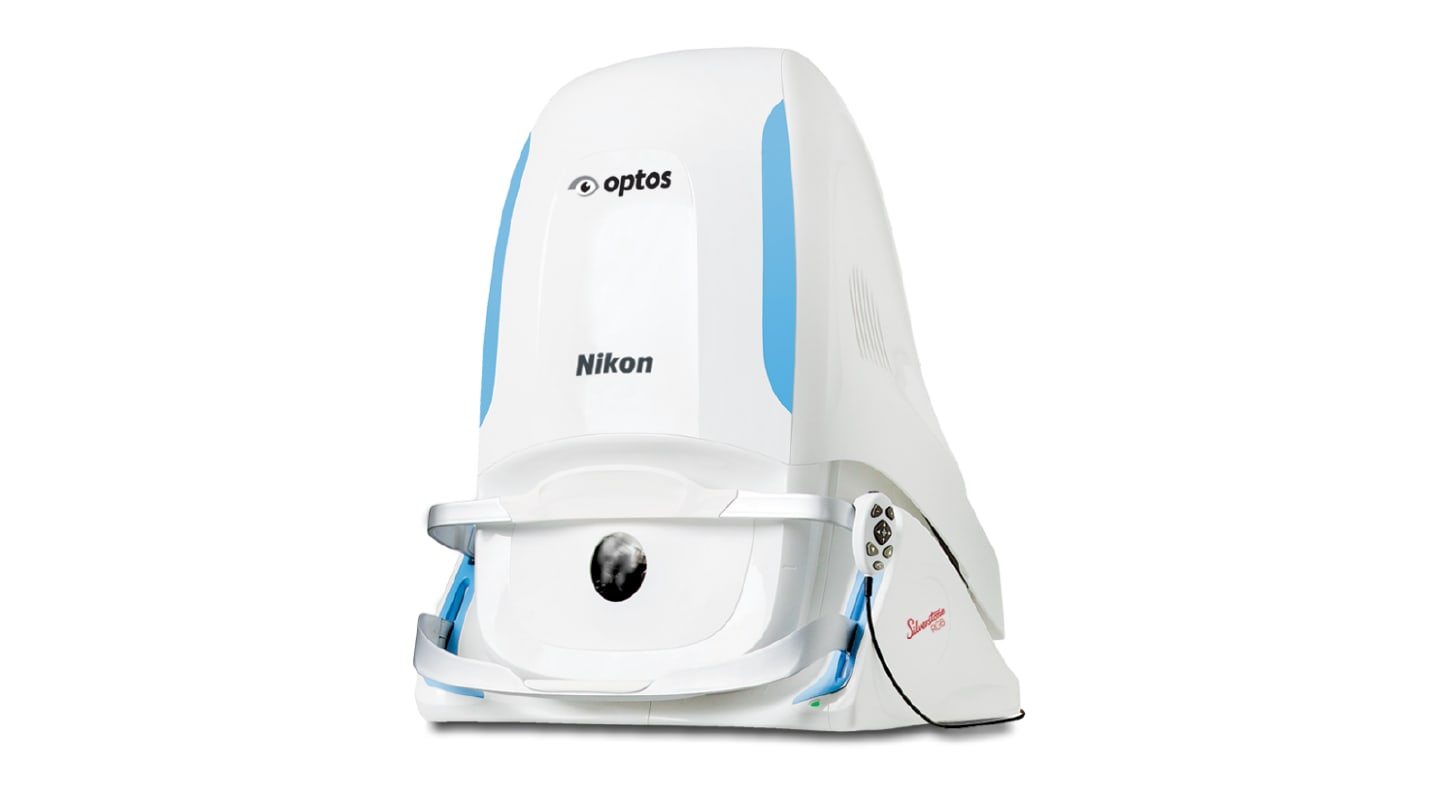
The Silverstone RGB system includes nine imaging modalities for surface to deep tissue imaging, functional and metabolic imaging, vascular imaging, and structural cross-sectional imaging, significantly broadening diagnostic capabilities.
It retains the ultra-widefield imaging and lesion-guided swept-source OCT from the existing Silverstone platform, ensuring continuity in clinical practice.
Clinician feedback emphasized the need for greater access to peripheral pathology, intuitive modality switching, and faster image capturing, which were critical in shaping the system's development.
Interpretation:
The integrated approach of the Silverstone RGB system supports comprehensive pathology assessment, enhancing the ability of clinicians to make informed decisions based on a complete view of retinal health.
Limitations:
The article does not provide specific data on the effectiveness or clinical outcomes of the Silverstone RGB system, which is crucial for evaluating its impact on patient care.
Conclusion:
The Silverstone RGB system aims to enhance retinal imaging capabilities while ensuring accessibility and efficiency in clinical practice, ultimately improving patient outcomes.
In an observational US target trial emulation, glucagon-like peptide-1 receptor agonist initiation was associated with about 3 to 4 more ischemic optic neuropathy cases per 10,000 patients over 18 months than two comparator drug classes.