Conexiant’s news site is now MDSpire News. Learn more
Advertisement
Multimodal Imaging in Inherited Retinal Disorders
As an increasing number of IRD patients are being identified and referred for genetic testing and counseling, clinicians would benefit from a practical and efficient framework for integrating multimodal imaging into their diagnoses and management. This review summarizes the utility of multimodal imaging across IRDs.
To summarize the utility of multimodal imaging techniques in the diagnosis and management of inherited retinal disorders (IRDs), emphasizing their significance in improving patient outcomes.
Approach:
Key Findings:
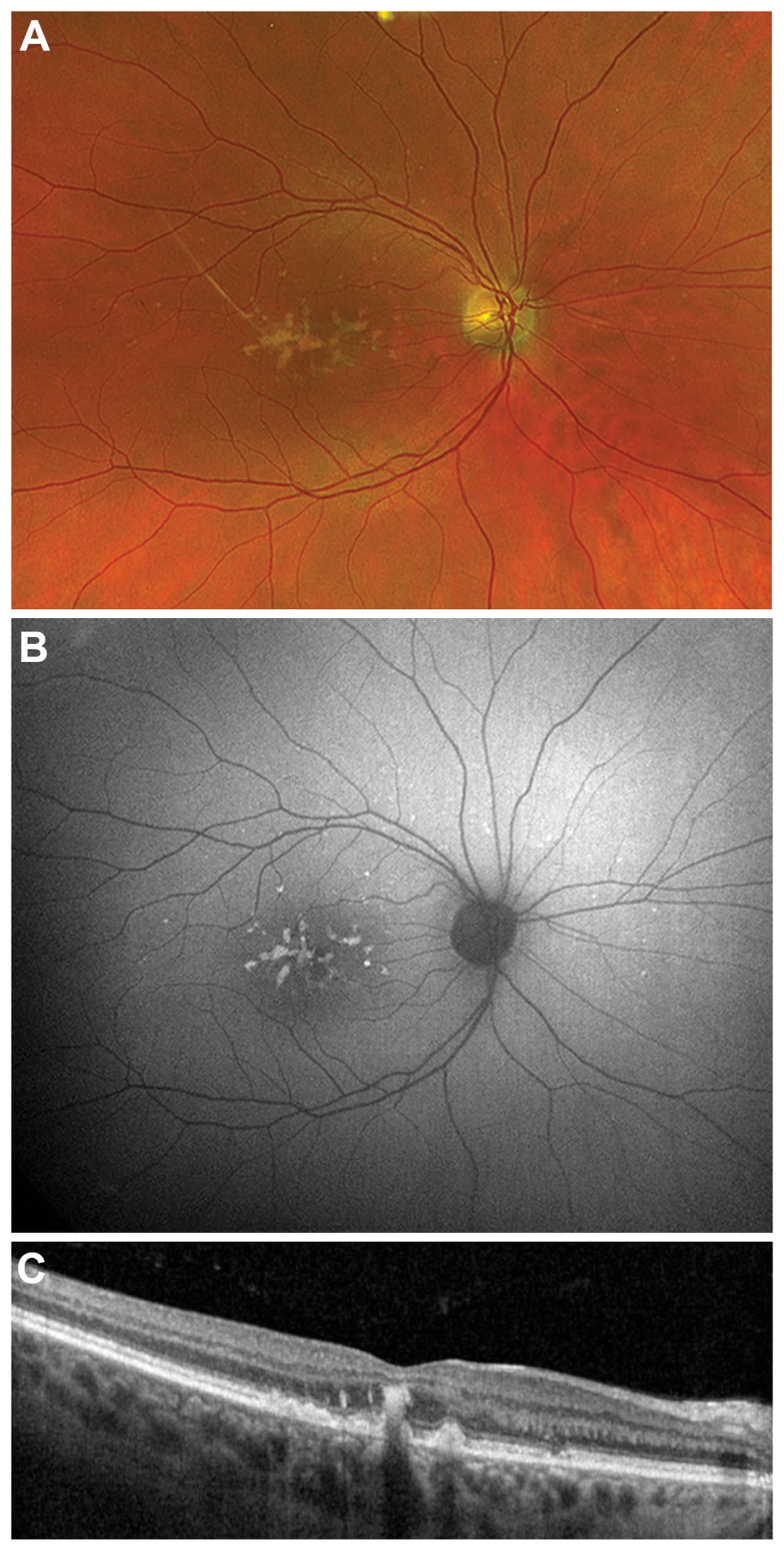
Fundus photography and ultrawidefield imaging are essential for documenting retinal features and detecting peripheral abnormalities, with specific examples of their clinical relevance.
Fundus autofluorescence (FAF) is crucial for early diagnosis and monitoring of IRDs, revealing abnormalities not visible on standard examination, supported by data from recent studies.
NIR-AF and quantitative autofluorescence (qAF) provide complementary information to conventional FAF, enhancing diagnostic accuracy, with examples of their application in clinical settings.
Interpretation:
Multimodal imaging significantly enhances the ability to diagnose, monitor, and understand the progression of inherited retinal disorders, facilitating better patient management and highlighting implications for future research.
Limitations:
Machine learning approaches for automated FAF interpretation face challenges due to image variability, but ongoing research is exploring solutions.
Current imaging techniques may not be universally applicable to all patient populations, particularly those with specific anatomical variations, necessitating further investigation.
Conclusion:
The integration of multimodal imaging into clinical practice is essential for improving the diagnosis and management of inherited retinal disorders, especially as gene-based therapies become more prevalent, underscoring the urgency of this integration.