A case report and literature review on imaging manifestations of immature teratoma in fetal oral cavity with concurrent intracranial abnormalities - Summary - MDSpire
Advertisement
A case report and literature review on imaging manifestations of immature teratoma in fetal oral cavity with concurrent intracranial abnormalities
To explore the imaging characteristics of immature teratoma in the fetal oral cavity and improve diagnostic and differential diagnostic capabilities for this condition.
Key Findings:
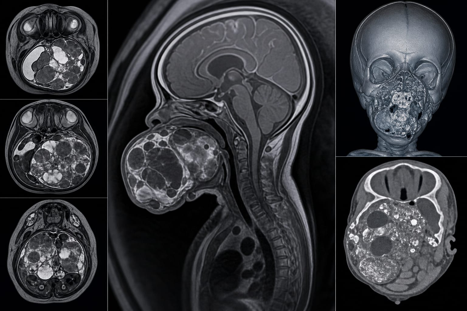
Imaging manifestations included a large solid-cystic mass in the fetal oropharynx, significant for differential diagnosis.
CT revealed heterogeneous density with cystic appearance and punctate calcifications, a feature more commonly associated with immature teratomas.
MRI showed irregular solid-cystic mass with specific signal characteristics correlating with histopathology, aiding in accurate diagnosis.
Interpretation:
Understanding the imaging features and associated intracranial abnormalities can significantly improve diagnostic proficiency for fetal oral cavity teratomas, impacting clinical decision-making.
Limitations:
The rarity of the condition limits the number of available cases for comprehensive analysis, potentially affecting the generalizability of findings.
Potential variability in imaging techniques and interpretations across different institutions may influence diagnostic outcomes.
Conclusion:
Multimodal imaging enhances prenatal diagnosis of immature teratomas, allowing differentiation from other oral masses and emphasizing the importance of evaluating concurrent intracranial abnormalities in clinical practice.