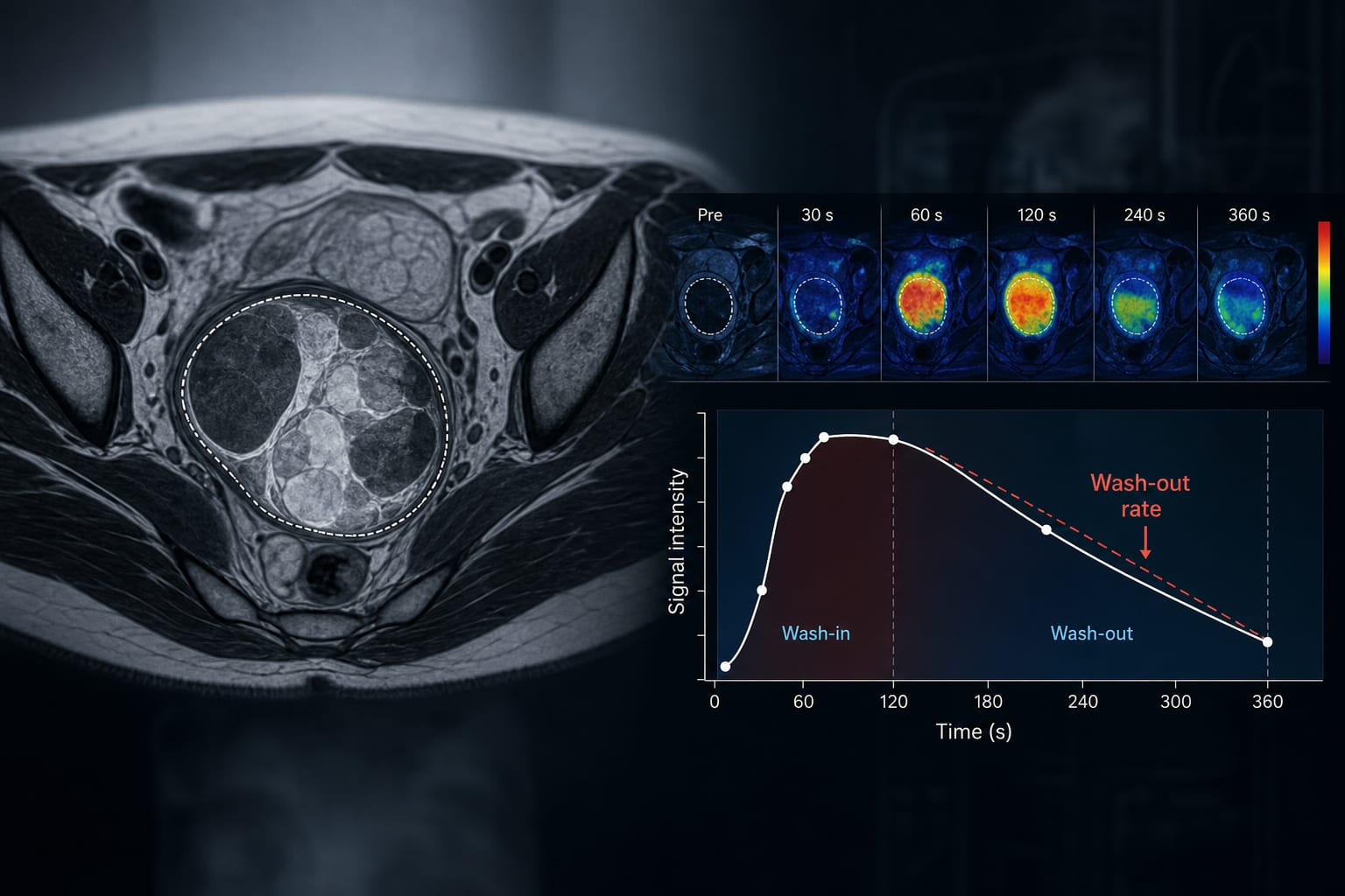
To assess the role of semiquantitative MR perfusion parameters, including wash-out rate, in differentiating benign from malignant ovarian masses, particularly focusing on their clinical implications.
Key Findings:
Significant correlation between pathological diagnosis and T2 signal intensity, ascites, and peritoneal deposits (p < 0.05).
Wash-out rate (WOR) and maximum absolute enhancement (SI max) significantly differentiated benign and malignant lesions (p < 0.05).
Cutoff value of WOR >6.03 provided accuracy of 86.2%, sensitivity of 82.4%, specificity of 90%, PPV of 93.3%, and NPV of 75%.
Cutoff value of SI max >1391.07 provided accuracy of 73.2%, sensitivity of 76.5%, specificity of 70%, PPV of 81.2%, and NPV of 63.6%.
Interpretation:
The addition of DCE MR Sequence to conventional MRI significantly enhances diagnostic accuracy for ovarian masses, with important implications for clinical practice.
Limitations:
Small sample size of 29 patients may limit the generalizability of the findings.
Exclusion of patients with simple cystic adnexal lesions or solid components <3 mm may affect the applicability of results.
Conclusion:
DCE MRI enhances the ability to differentiate malignant from benign ovarian masses, providing critical information for surgical planning and patient management.
by Heba Sabet, Moawia Gameraddin, Fahad H. Alhazmi, Walaa Alsharif, Nagham N. Omar, Ahmed Sobh, Sultan A. Alshoabi, Awadia Gareeballah, Maisa Elzaki, Hadeel A. Ghunaim, Awatif M. Omer, Amel F. H. Alzain, Reem M. Elkady