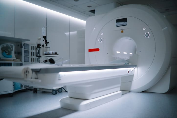
To investigate anatomical variations regarding the number of meniscal root insertions and their corresponding insertion locations in asymptomatic subjects using 7-T MRI.
Key Findings:
All scanned knees exhibited intact meniscal root insertions.
Meniscal tears were observed in six knees, primarily affecting the pars intermedia and posterior meniscal horn.
The signal intensity of the menisci was hypointense with fine horizontal hyperintense striations.
Interpretation:
High-resolution 7-T MRI effectively visualizes meniscal anatomy and variations, enhancing diagnostic confidence in identifying meniscal root insertions.
Limitations:
Study limited to asymptomatic individuals, which may not represent the general population.
Exclusion of participants with claustrophobia or metal implants may limit generalizability.
Conclusion:
The study highlights the potential of 7-T MRI in elucidating meniscal anatomy and variants, which could improve surgical planning and outcomes.