Autoimmune GFAP astrocytopathy with eosinophils on cerebrospinal fluid cytology and isolated spinal cord lesions on MRI: a case report - Summary - MDSpire
Advertisement
Autoimmune GFAP astrocytopathy with eosinophils on cerebrospinal fluid cytology and isolated spinal cord lesions on MRI: a case report
To present a case of autoimmune GFAP astrocytopathy characterized by eosinophils in cerebrospinal fluid (CSF) cytology and isolated thoracic spinal cord lesions on MRI, highlighting the significance of eosinophils in the diagnostic process.
Key Findings:
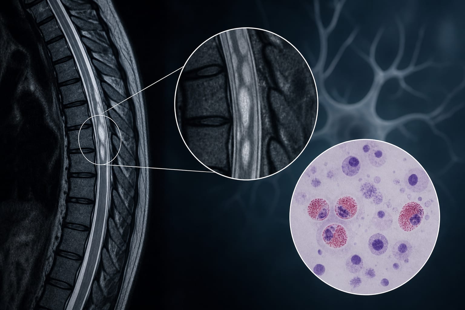
The patient had elevated opening pressure, pleocytosis, increased protein, and 10% eosinophils in CSF.
Brain MRI was unremarkable, while spinal MRI revealed discontinuous patchy long-segment intramedullary lesions.
CSF GFAP-IgG was positive at a titer of 1:32, while serum GFAP-IgG was negative.
Infectious studies, including CSF culture and metagenomic next-generation sequencing, were negative.
The patient improved significantly after treatment with high-dose intravenous methylprednisolone.
Interpretation:
Isolated spinal cord lesions on MRI may indicate autoimmune GFAP astrocytopathy, even in the absence of brain abnormalities. Eosinophils in CSF may suggest a unique inflammatory profile, warranting further investigation.
Limitations:
The case is a single patient report, limiting generalizability.
Eosinophils in CSF are rare and may complicate the diagnostic process, potentially leading to misdiagnosis.
Conclusion:
The presence of eosinophils in CSF and isolated spinal cord lesions should prompt consideration of autoimmune GFAP astrocytopathy, highlighting the need for further research on the role of eosinophils in this condition.
A fatal NEJM case highlights how invasive meningococcal disease—particularly serogroup W—can present without rash, masquerade as gastrointestinal illness, and rapidly progress to shock and DIC.