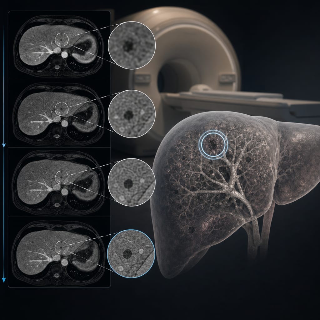
To investigate the clinical utility of fully automated 3D organ segmentation in assessing hepatic steatosis on pre-contrast and post-contrast CT images, using MRS-PDFF as the reference standard for accuracy.
Key Findings:
Volumetric CT attenuation measurements may provide a more comprehensive representation of liver characteristics compared to ROI-based measurements, suggesting a shift in assessment techniques.
CT-based hepatic steatosis assessment relies on differences in X-ray absorption between triglycerides and normal liver tissue, highlighting the need for accurate measurement techniques.
Automated segmentation techniques can efficiently and objectively obtain organ volumes and radiomics features, potentially transforming clinical practice.
Interpretation:
The study suggests that automated 3D organ segmentation could enhance the assessment of hepatic steatosis, potentially offering a non-invasive alternative to traditional methods.
Limitations:
Limited data on the accuracy of volumetric CT attenuation measurements compared to ROI-based measurements, which may affect the reliability of the findings.
The study's retrospective nature may introduce selection bias, necessitating caution in generalizing results.
Conclusion:
Fully automated 3D organ segmentation shows promise for improving the assessment of hepatic steatosis using CT, warranting further investigation to validate its clinical utility.