To present a case of glomus tumor of the stomach and discuss its diagnostic and therapeutic approaches, highlighting its rarity and potential for misdiagnosis.
Key Findings:
Cytopathology revealed uniform, round epithelioid cells positive for smooth muscle actin, with negative markers for desmin and others.
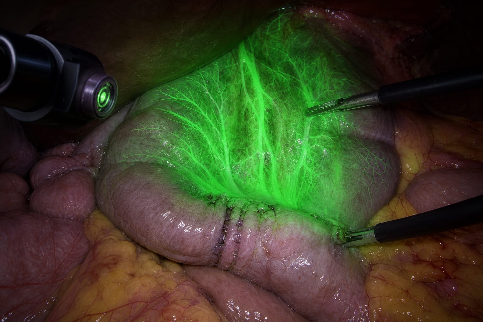
CT scan confirmed a hyperdense lesion without lymphadenomegaly or metastasis, measuring approximately 14 mm.
Histological examination confirmed the diagnosis with clear surgical margins, detailing the tumor's characteristics.
Interpretation:
Glomus tumors of the stomach are rare, often misdiagnosed as gastric cancer or GIST, and can be effectively diagnosed using EUS-FNA and treated with laparoscopic resection.
Limitations:
Lack of clear guidelines for optimal therapy of glomus tumors, which may lead to varied treatment approaches.
Potential for misdiagnosis as gastric cancer or GIST, highlighting the need for accurate diagnostic methods.
Conclusion:
EUS-FNA is the gold standard for diagnosing glomus tumors, and laparoscopic wedge resection is a reasonable therapeutic approach due to their benign nature, emphasizing the importance of accurate diagnosis.