To enhance understanding of the imaging features of breast epithelial–myoepithelial carcinoma (EMC) through a case study and literature review.
Approach:
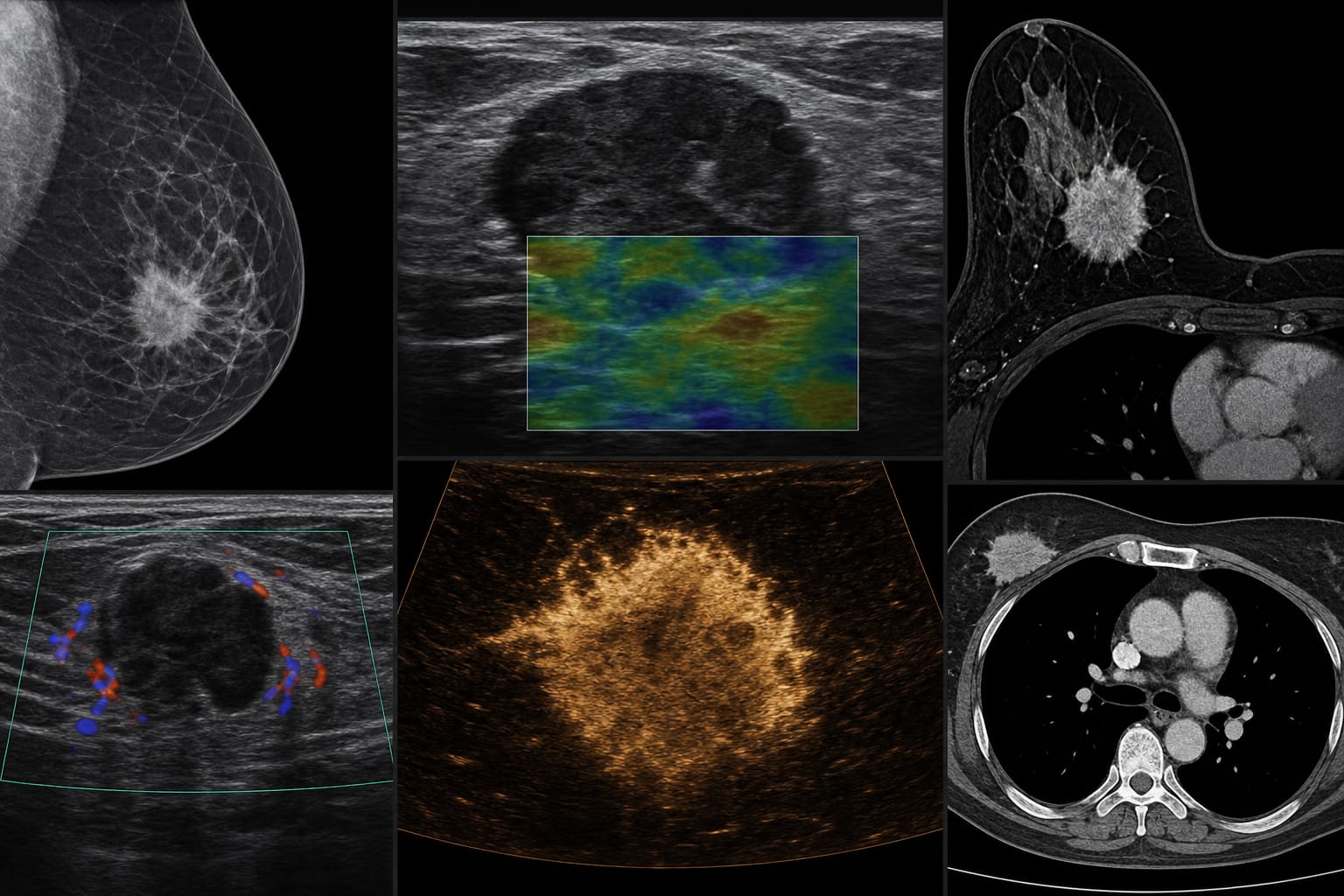
Imaging Techniques: The study presents and analyzes imaging findings in correlation with pathological results, highlighting the significance of SWE and CEUS in assessing breast lesions.
Key Findings:
Breast EMC accounts for less than 0.5% of all breast tumors.
Imaging characteristics of EMC are non-specific and can overlap with other breast conditions.
SWE demonstrated stiffness heterogeneity, while CEUS showed intratumoral perfusion heterogeneity.
Interpretation:
The case highlights the potential role of multimodal imaging, particularly SWE and CEUS, in the preoperative assessment of breast EMC.
Limitations:
The rarity of breast EMC limits the generalizability of findings to broader populations.
Existing literature on imaging characteristics of EMC is limited and primarily consists of single-case reports.
Conclusion:
This case contributes to the limited imaging literature on breast EMC and illustrates advanced ultrasound techniques in evaluating rare breast tumors.