To report a neonatal case of horseshoe lung (HL) complicated with scimitar syndrome, VACTERL association, and intestinal malrotation, enhancing understanding of HL and its associated anomalies.
Approach:
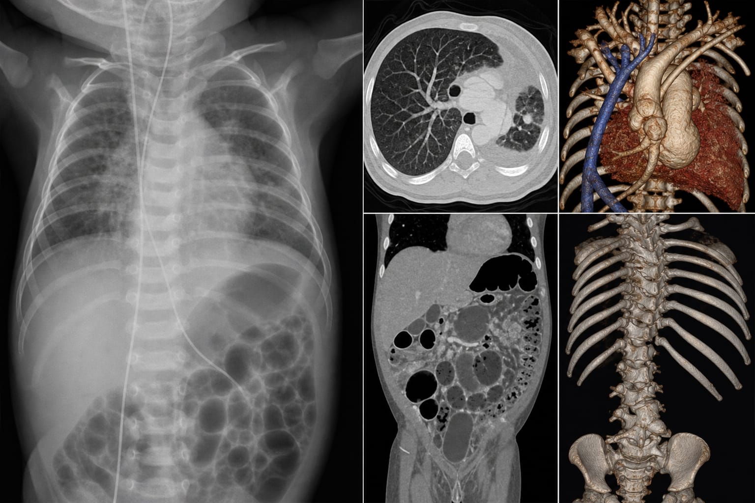
Case Description: A premature female neonate presented with respiratory distress and multiple congenital anomalies, including HL, scimitar syndrome, and intestinal malrotation.
Imaging Techniques: Multiple imaging modalities, including echocardiography, abdominal ultrasonography, thoracoabdominal x-ray, and chest CTA, were utilized for comprehensive evaluation.
Key Findings:
The patient exhibited horseshoe lung with abnormal bronchial branching and pulmonary hypoplasia, identified through various imaging modalities.
Associated anomalies included scimitar syndrome, complex cardiovascular malformations, duodenal obstruction, and multiple vertebral anomalies.
Surgery was performed to relieve gastrointestinal obstruction, revealing annular pancreatic tissue.
Interpretation:
The coexistence of HL with various systemic anomalies presents challenges for clinical diagnosis and management.
Limitations:
The case study is based on a single patient, limiting generalizability to broader populations.
Long-term outcomes and prognosis were not assessed in the report.
Conclusion:
This case highlights the complexity of diagnosing and managing HL in the context of multiple congenital anomalies.