To compile current evidence regarding key imaging biomarkers pertinent to the individualized management of diabetic macular edema (DME) and explore their incorporation into an imaging phenotype-based methodology for treatment selection.
Approach:
Key Findings:
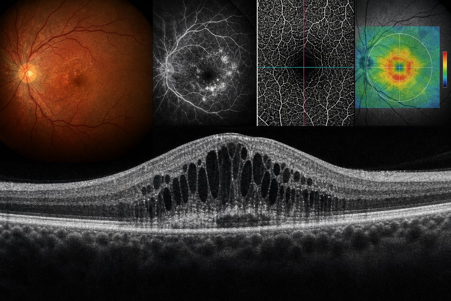
Markers of disease activity include intraretinal and subretinal fluid along with hyperreflective foci.
Predictors of visual prognosis consist of disorganization of the retinal inner layers, photoreceptor injury, and retinal perfusion deficits.
DME can be classified into five clinical phenotypes: leakage-dominant, inflammatory, tractional, focal-treatment, and poor-prognosis phenotypes.
Interpretation:
The integration of imaging biomarkers into a proposed, hypothesis-generating decision-support framework aims to align therapeutic approaches with underlying pathogenic mechanisms in DME.
Limitations:
Variability in imaging acquisition protocols and biomarker specifications across studies.
Limited high-level evidence for some emerging biomarkers.
Conclusion:
Future advancements in automated biomarker quantification and AI-assisted image analysis may enhance precision in DME phenotyping and facilitate individualized disease management.
Presenting results from the DME AWARE Delphi Study at the Association for Research in Vision and Ophthalmology (ARVO) meeting in Denver, Baruch D. Kuppermann, MD, PhD, director of the Gavin Herbert Eye Institute at the University of California, Irvine, described a set of consensus findings that point to unmet needs across the continuum of DME management, with particular emphasis on early intervention and noninvasive treatment options (Figure 1).