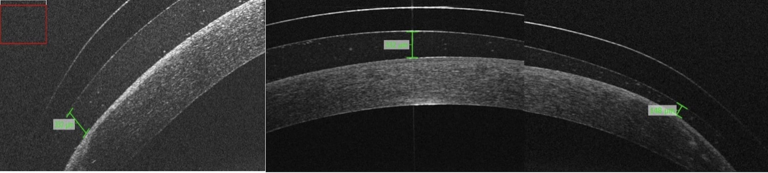
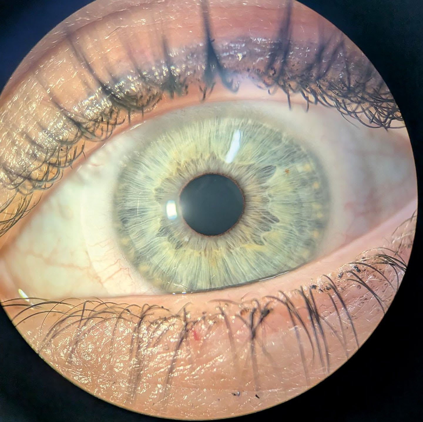
To illustrate the effectiveness of profilometry-guided scleral lens design in managing keratoconus and its complications, as demonstrated in a case study.
Approach:
Case Description: A 45-year-old male with keratoconus and a penetrating keratoplasty in his right eye experienced complications from poorly designed scleral lenses, including a pyogenic granuloma, and reported constant redness and irritation.
Lens Fitting: Scleral profilometry was performed, and customized Latitude freeform lenses were ordered to match the scleral surface shape, significantly reducing compression over the granuloma.
Follow-Up: The patient reported improved comfort and vision after lens adjustments, achieving 20/20 vision in both eyes, with significant reduction in granuloma size and complete resolution over time.
Key Findings:
The patient achieved 20/20 vision in both eyes after lens adjustments.
Profilometry-guided lens design reduced conjunctival compression and resolved the pyogenic granuloma.
Sequential lens modifications were necessary as the ocular surface changed.
Interpretation:
The case highlights the importance of individualized scleral lens design to accommodate the unique topography of the eye in keratoconus.
Limitations:
The study is based on a single case report, limiting generalizability.
Further research is needed to evaluate long-term outcomes of profilometry-guided scleral lenses.
Conclusion:
Profilometry-guided scleral lens design led to the complete resolution of the pyogenic granuloma and improved visual acuity and comfort for the patient.