To describe the cuneus vein, a previously unreported cortical venous structure, and propose techniques for its preservation during posterior interhemispheric approaches, highlighting its significance in neurosurgery.
Key Findings:
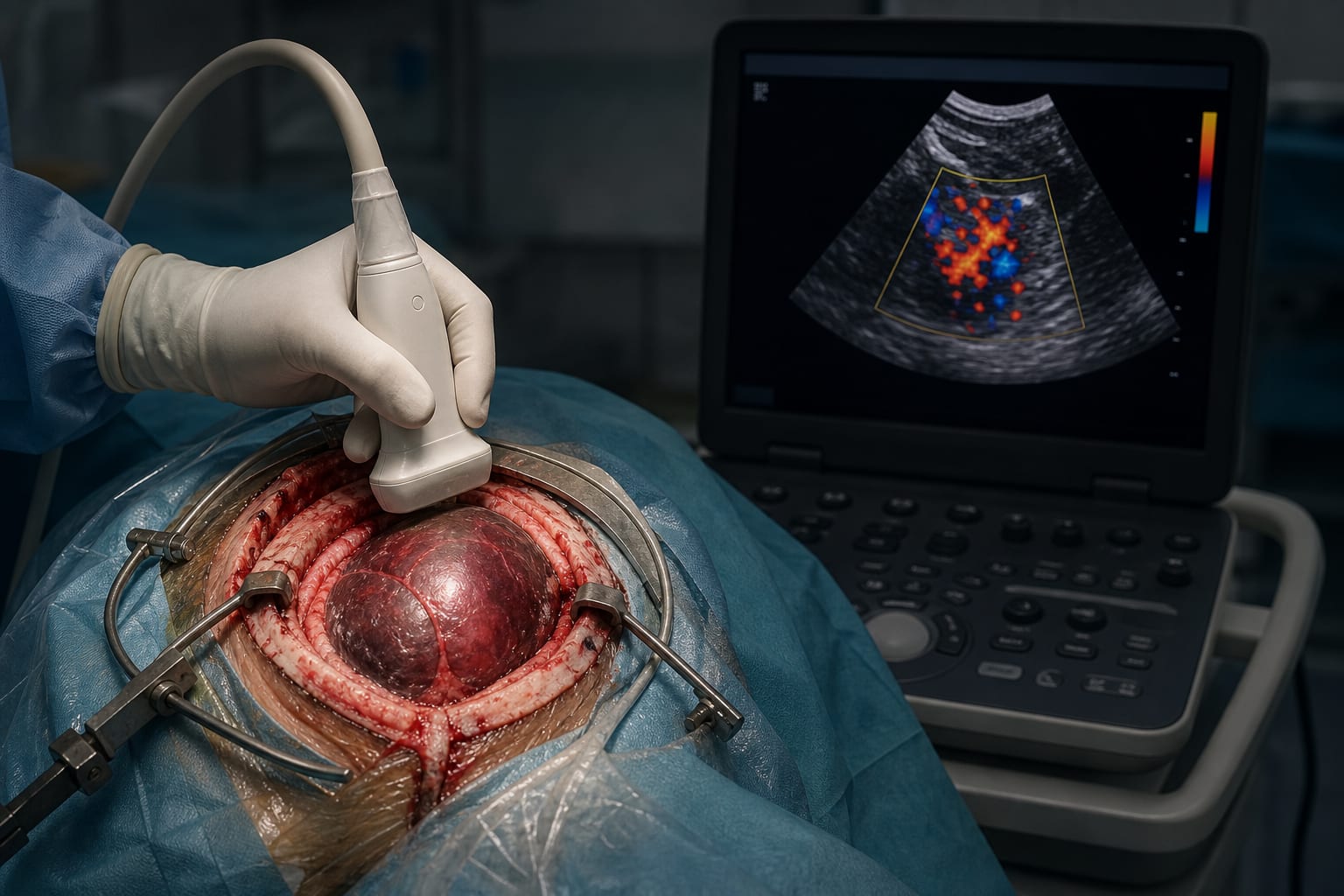
Preservation of venous flow is crucial in neurosurgery.
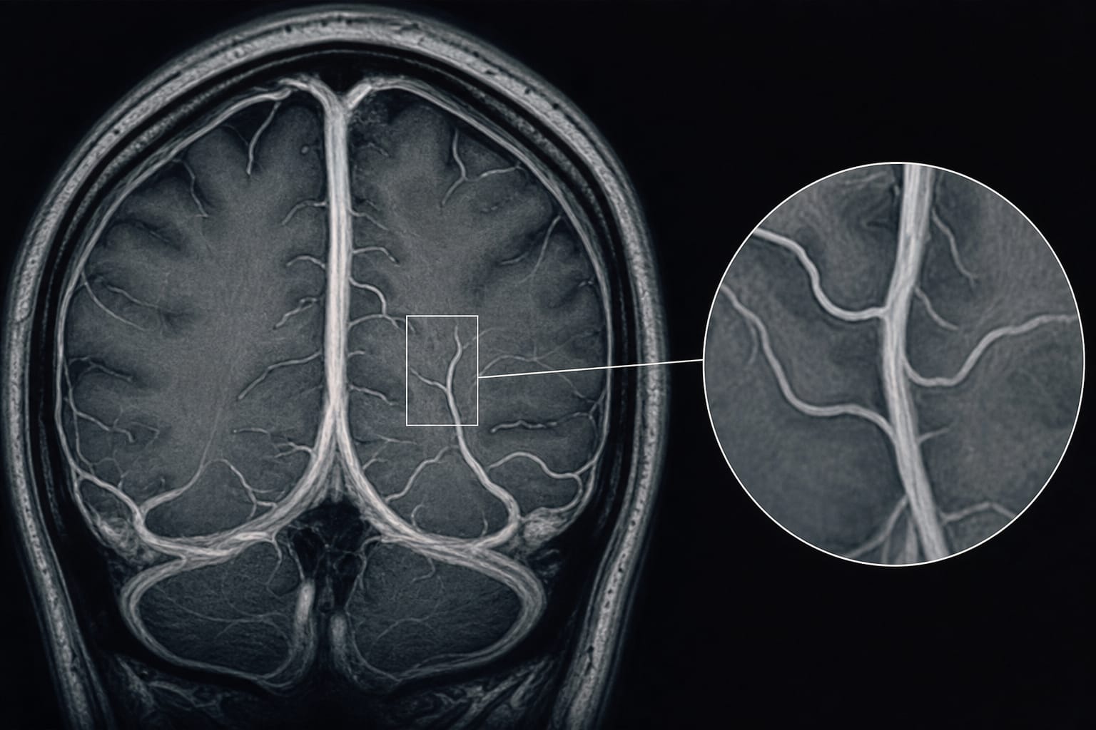
The cuneus vein was identified intraoperatively but not visualized in preoperative MRI scans, indicating potential imaging limitations.
The study's methodology has notable limitations, including potential bias from a single operator and institution.
Interpretation:
The inability to visualize the cuneus vein in preoperative MRI raises questions about its identification and the validity of the imaging methods used, suggesting a need for improved imaging techniques.
Limitations:
Retrospective design introduces potential bias.
Study confined to a single operator and institution, limiting generalizability.
Small number of cases weakens conclusions.
Lack of detailed methodology and results for retrospective MRI analysis, including scanner type and imaging protocols.
Conclusion:
The validity of the cuneus vein as a novel anatomical structure remains debatable, necessitating further validation using appropriate imaging techniques and methodologies.