To establish a whole, large-format brain slice model that preserves the complex cellular architecture of the brain, enabling more effective glioblastoma research.
Approach:
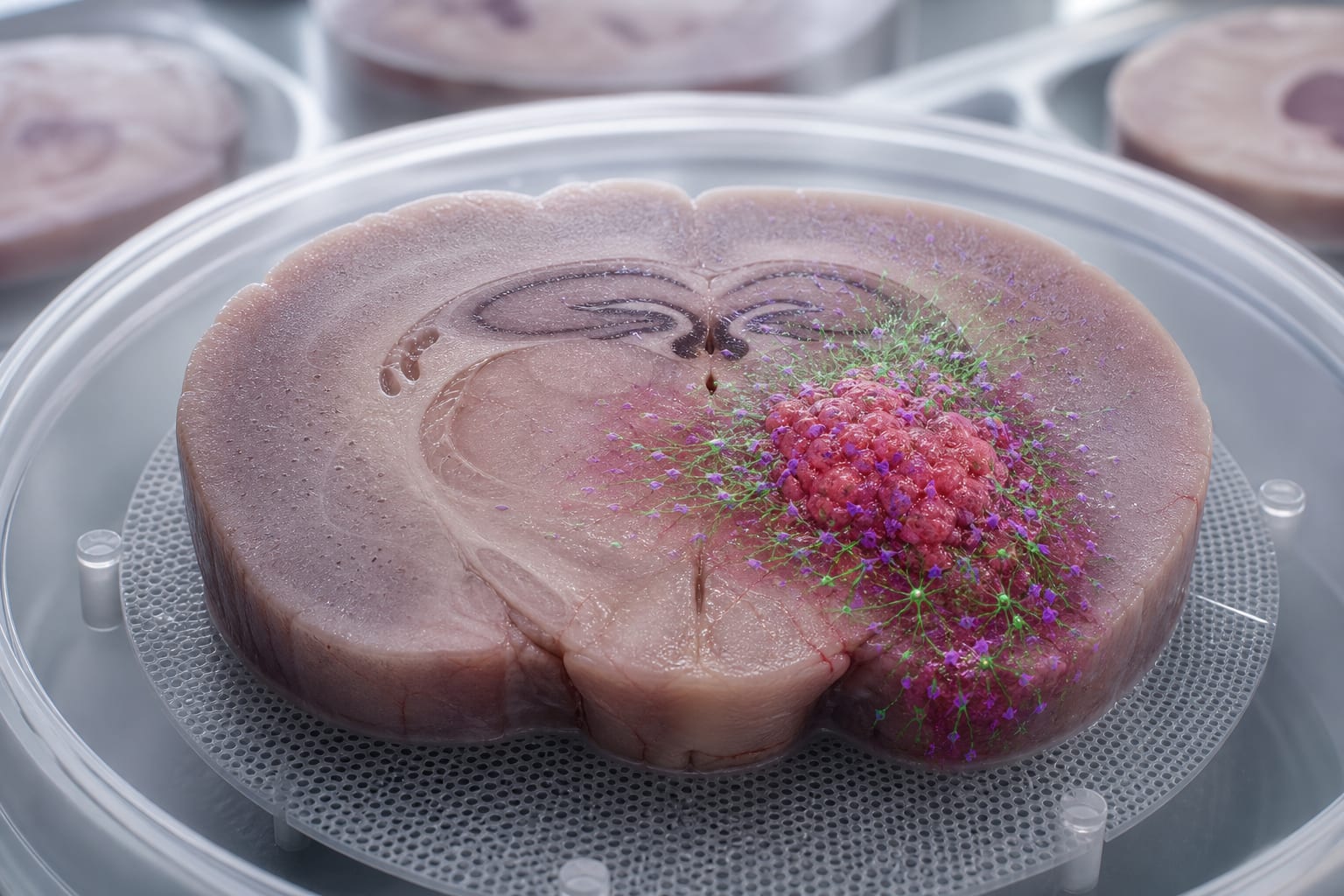
Model Development: Created a whole murine brain slice model using 6-9-day-old postnatal mice, allowing for multiple contiguous slices to be sectioned and maintained in vitro.
Tumor Cell Integration: Co-cultured rat 9L/lacZ GBM cell spheroids with the brain slices to simulate tumor growth and invasion.
Viability Assessment: Utilized AlamarBlue assay and live/dead staining to visualize and quantify slice viability and structural integrity over time, ensuring accurate assessment of the model's health.
Key Findings:
Brain slices remained viable and stable for at least 7 days, with potential for extended culture duration as assessed by AlamarBlue and live/dead staining.
Co-culture with GBM spheroids led to a slight decrease in slice viability due to tumor invasion.
The model preserves organotypic brain and tumor structures, facilitating the study of GBM biology.
Interpretation:
The developed ex vivo whole-brain slice GBM model provides a platform for investigating glioblastoma interactions with the tumor microenvironment.
Limitations:
The model's viability gradually decreases after one week, which may limit long-term studies.
Potential ethical considerations regarding the use of animal-derived tissues should be addressed.
Conclusion:
This model serves as a valuable tool in brain cancer research, effectively bridging the gap between traditional 2D cultures and complex in vivo models.