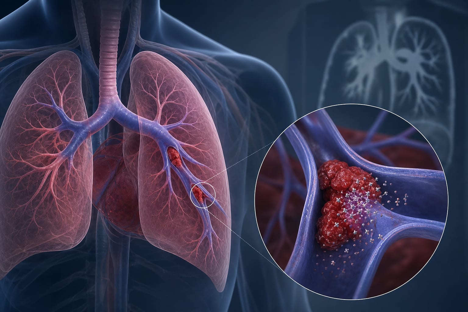
To highlight the etiology, differential diagnosis, and management of pulmonary artery intimal membrane hemorrhage (PA-IMH) in patients presenting with acute chest and back pain and hypoxemia.
Approach:
Etiology: PA-IMH represents a special type of PA dissection associated with severe pulmonary hypertension (mPAP > 50 mmHg), most commonly attributable to congenital heart disease. Chronic pressure overload enhances wall tension, while jet direction through patent ductus arteriosus (PDA) increases local shear stress on left PA, predisposing to localized intimal injury and subsequent unilateral IMH/dissection. Hypoxemia results from mPAP exceeding mean systemic pressure with consequent right-to-left shunting—i.e., Eisenmenger’s syndrome, accounting for 0.9% of PDA cases.
Differential Diagnosis: In patients presenting with acute chest/back pain and hypoxemia, pulmonary embolism (PE) is characterized by irregular intraluminal defects, distinct from the smooth, continuous wall thickening of PA-IMH. Other entities—acute coronary syndrome (ACS) (wall motion abnormalities), aortic dissection (AAS) (aortic intimal flap), and pericardial effusion (AP)—are readily distinguished by transthoracic echocardiography (TTE) and rarely cause hypoxemia.
Management: A conservative, physiology-directed strategy is generally preferred for PA-IMH. Pharmacotherapy targeting pulmonary hypertension (e.g., prostacyclin analogs) may attenuate IMH progression. Close imaging surveillance is advised, with surgery reserved for overt PA dissection given high mortality.
Key Findings:
PA-IMH is a rare but potentially fatal complication of severe pulmonary hypertension.
Transthoracic echocardiography (TTE) enables timely bedside detection and differential diagnosis in acute care settings.
Interpretation:
In cases of prolonged severe pulmonary hypertension, sudden chest/back pain may indicate PA-IMH.
Conclusion:
Timely diagnosis and management are crucial for improving outcomes in patients with PA-IMH.