To characterize the MRI features of cervical metastatic lymph nodes in differentiated thyroid carcinoma (DTC) and provide evidence for identifying these nodes prior to radioactive iodine (¹³¹I) therapy.
Approach:
Key Findings:
Cervical US detected lymph node metastasis in 15.6% of cases.
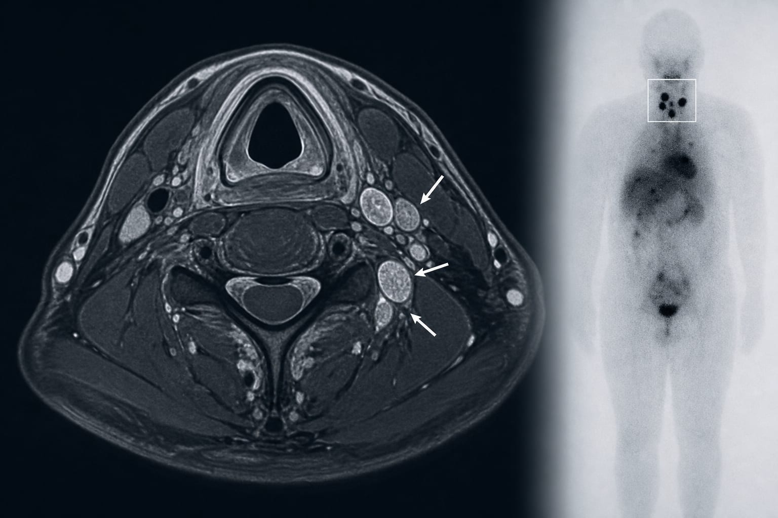
A total of 58 radioiodine-avid lymph nodes were identified across cervical levels II–VI and the supraclavicular region.
Most radioiodine-avid lymph nodes exhibited hyperintensity on T1- and T2-weighted sequences.
65.5% of nodes were round to ovoid, and 32.8% were irregular in shape.
69.0% of nodes had ill-defined margins, 91.4% lacked a lymphatic hilum, and 77.6% were adjacent to vascular structures.
In 96.9% (31/32) of patients, at least 4 lymph nodes were visualized on MRI.
Interpretation:
MRI features of cervical lymph nodes with radioiodine uptake often show overlapping characteristics with radioiodine-negative nodes, necessitating comprehensive assessment for accurate pre-therapeutic evaluation.
Limitations:
The study is retrospective and based on a limited sample size of 32 patients, which may affect the generalizability of the findings.
Imaging features of radioiodine-negative lymph nodes may overlap with those of radioiodine-avid nodes.
Conclusion:
Cervical lymph nodes with radioiodine uptake frequently present with specific MRI characteristics, but a multimodal imaging approach is essential for accurate assessment before ¹³¹I therapy.