To evaluate early anatomical changes and intraocular inflammation (IOI) following the switch to brolucizumab in patients with neovascular age-related macular degeneration (nAMD) who were refractory to aflibercept or ranibizumab.
Approach:
Study Design: Retrospective observational study analyzing data from 23 patients switched to brolucizumab.
Parameters Measured: Central foveal retinal thickness (CRT), pigment epithelial detachment volume, central choroidal thickness, and central retinal volume (CRV) were assessed.
Intraocular Inflammation Assessment: IOI was evaluated through clinical observation and OCT imaging.
Key Findings:
Significant decrease in CRT (from 247 to 227 μm, P < 0.001).
Decrease in pigment epithelial detachment volume (from 0.18 to 0.11 mm³, P = 0.04).
Increase in central choroidal thickness (from 159 to 168 μm, P = 0.018).
No significant change in central retinal volume (from 9.80 to 9.70 mm³, P = 0.111).
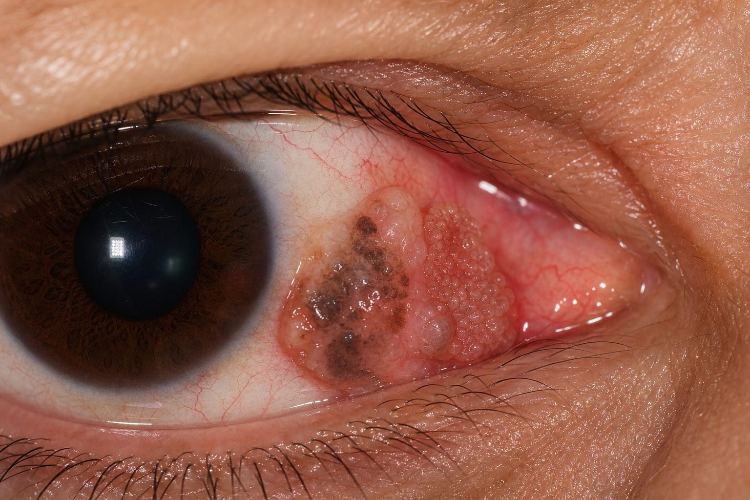
Three eyes exhibited IOI, with two showing mild anterior chamber inflammation and one exhibiting vasculitis obliterans.
Interpretation:
Limitations:
Small sample size of 23 patients.
Retrospective design may limit the ability to draw definitive conclusions.
Conclusion:
Switch to brolucizumab therapy resulted in early anatomical changes.