To evaluate the effectiveness of a retinal imaging-based model in detecting coronary artery disease (CAD) when combined with specific clinical risk factors such as age, sex, and lipid levels.
Approach:
Key Findings:
Combined model achieved AUROC of 0.802, sensitivity of 0.797, and specificity of 0.679.
Clinical risk factors alone had AUROC of 0.748; retinal data alone had AUROC of 0.694.
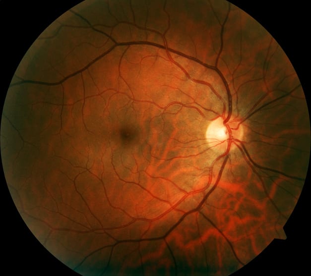
Independent retinal parameters associated with CAD included fractal dimension, vessel density, optic disc axis ratio, and optic disc-to-macula distance, with clear definitions of each parameter.
Interpretation:
The retinal microvasculature may serve as a noninvasive indicator of systemic vascular health, suggesting significant potential for enhancing cardiovascular risk assessment in clinical settings.
Limitations:
Retrospective, cross-sectional, single-center design limits causal inference and generalizability.
Cohort consisted of high-risk patients referred for angiography, not a general screening population.
Predominantly Han Chinese population and lack of broader biomarker incorporation limit applicability.
Conclusion:
Retinal vascular phenotyping may enhance traditional CAD risk assessment, particularly when combined with established risk factors, but further validation in diverse populations is necessary.