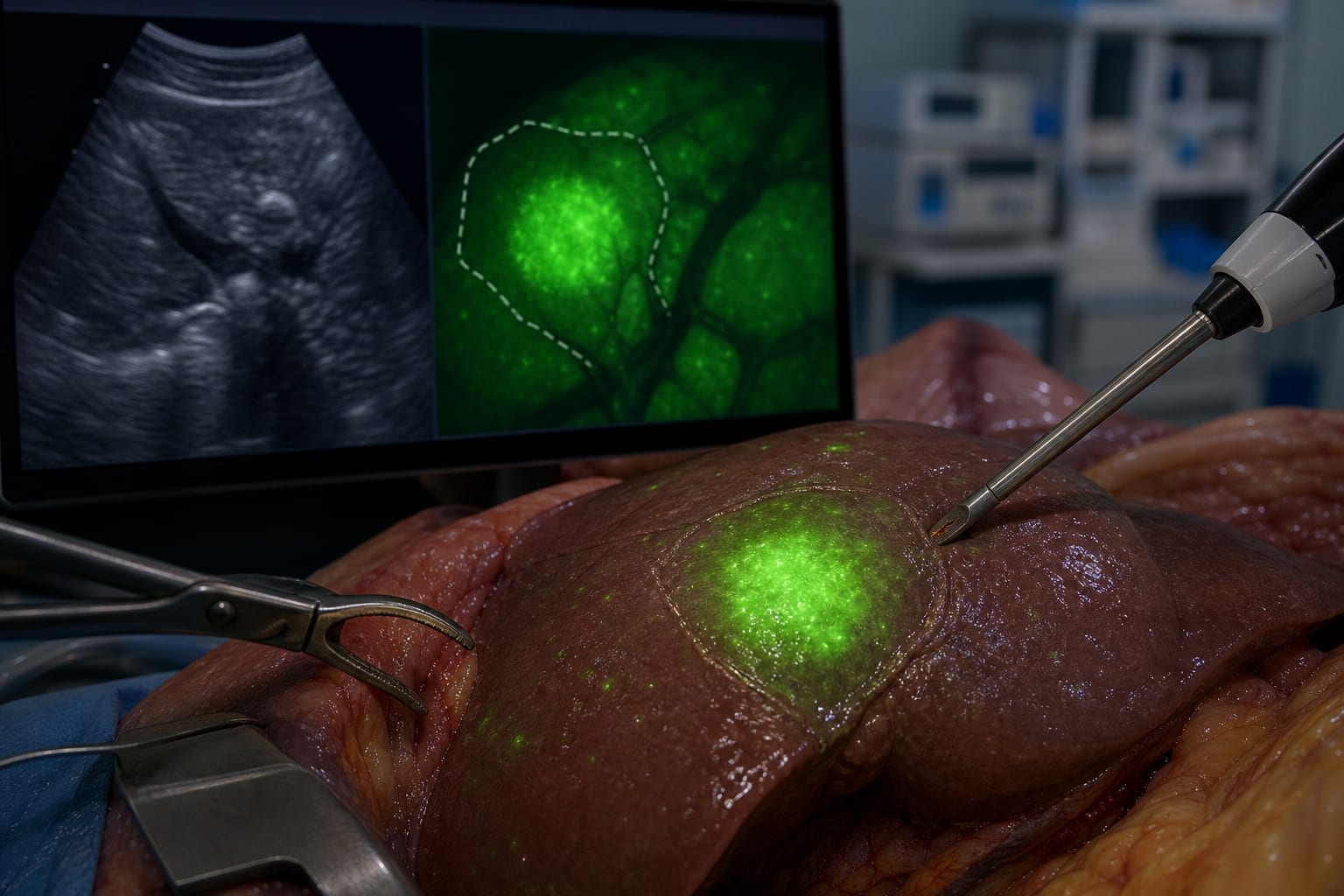
To describe the use of indocyanine green (ICG) fluorescence guidance combined with intraoperative ultrasonography (IOUS) for laparoscopic parenchyma-sparing excision of selected perivascular focal nodular hyperplasia (FNH).
Approach:
Key Findings:
Both patients underwent successful wedge excision with negative surgical margins.
Estimated blood loss was 50 mL and 20 mL for the two cases, respectively.
Incidental punctate hyperfluorescent foci were observed; one showed benign hyperplastic changes on histology.
Interpretation:
Remove unsupported conclusions about clinical value.
Limitations:
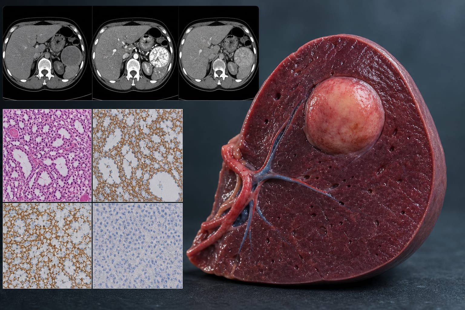
The absence of hepatobiliary phase MRI before surgery limited preoperative diagnostic workup in Case 1.
The clinical significance of incidental hyperfluorescent foci remains uncertain.
Conclusion:
Revise to reflect only the findings stated in the source without implications.