Case Report: Progressive acquired ileal mesenteric venous malformation after right hemicolectomy: serial imaging evolution leading to delayed gastrointestinal bleeding - Summary - MDSpire
Advertisement
Case Report: Progressive acquired ileal mesenteric venous malformation after right hemicolectomy: serial imaging evolution leading to delayed gastrointestinal bleeding
To report a rare case of progressive acquired ileal mesenteric venous malformation developing after right hemicolectomy and to highlight its imaging findings and management.
Approach:
Case Presentation: A 50-year-old woman presented with recurrent painless hematochezia and anemia four years after right hemicolectomy for colon cancer. Imaging revealed dilated veins in the ileal mesentery.
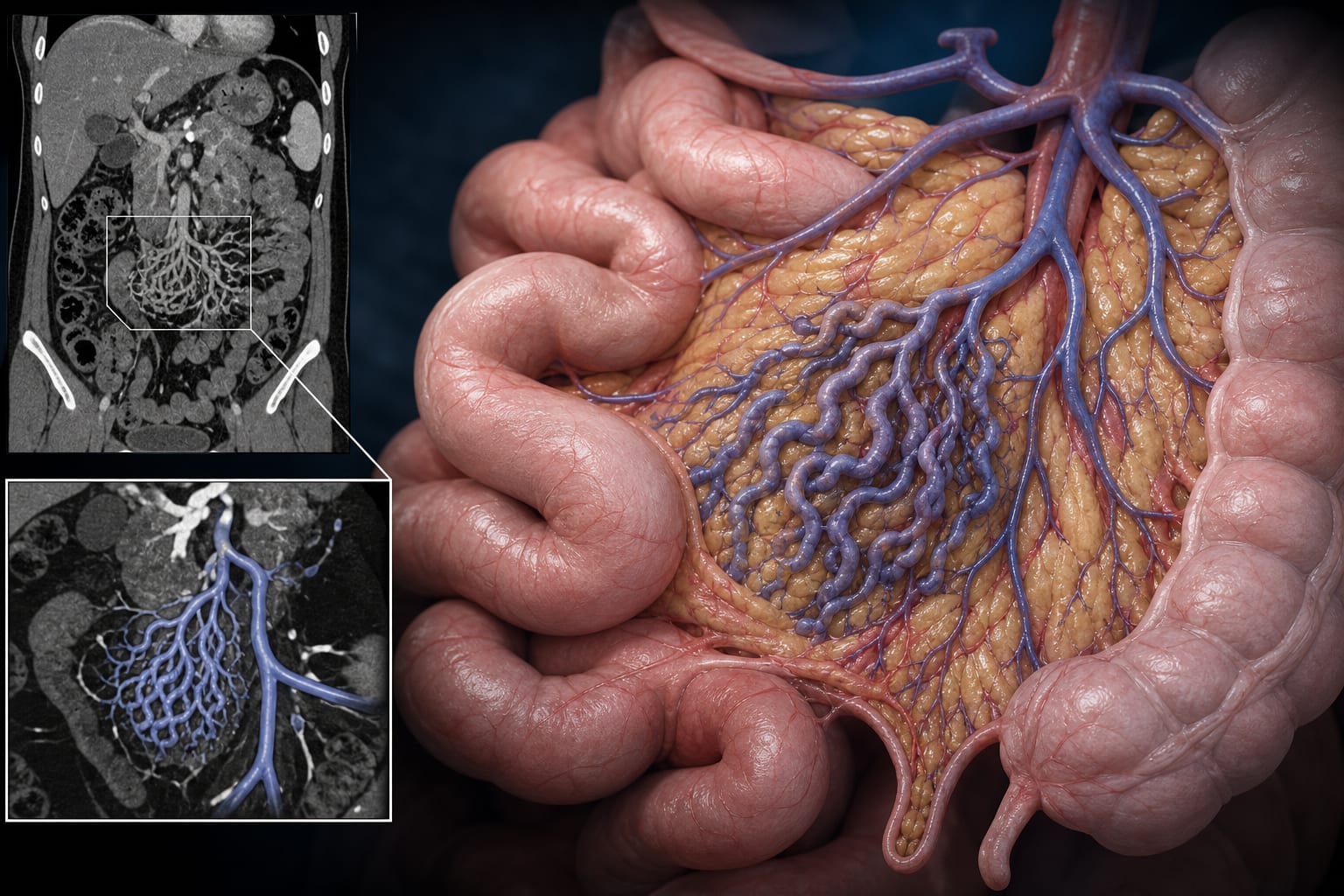
Imaging Findings: Contrast-enhanced CT angiography showed clusters of tortuous veins with a caput medusae appearance, draining into the superior mesenteric vein and communicating with the lumbar vein.
Surgical Intervention: Exploratory surgery confirmed a localized venous plexus, which was treated with targeted ligation and suturing, resulting in complete resolution of bleeding.
Key Findings:
Acquired ileal mesenteric venous malformation can develop years after intestinal surgery.
Imaging demonstrated progressive evolution of the venous malformation over time.
Surgical intervention led to complete resolution of gastrointestinal bleeding.
Interpretation:
Acquired ileal mesenteric venous malformations should be considered in patients with unexplained delayed gastrointestinal bleeding post-surgery, as they may result from postoperative venous outflow alterations.
Limitations:
The case is based on a single patient, limiting generalizability.
Long-term follow-up data are limited to 58 months post-surgery.
Conclusion:
Acquired ileal mesenteric venous malformations may facilitate timely diagnosis and surgical management in similar cases.