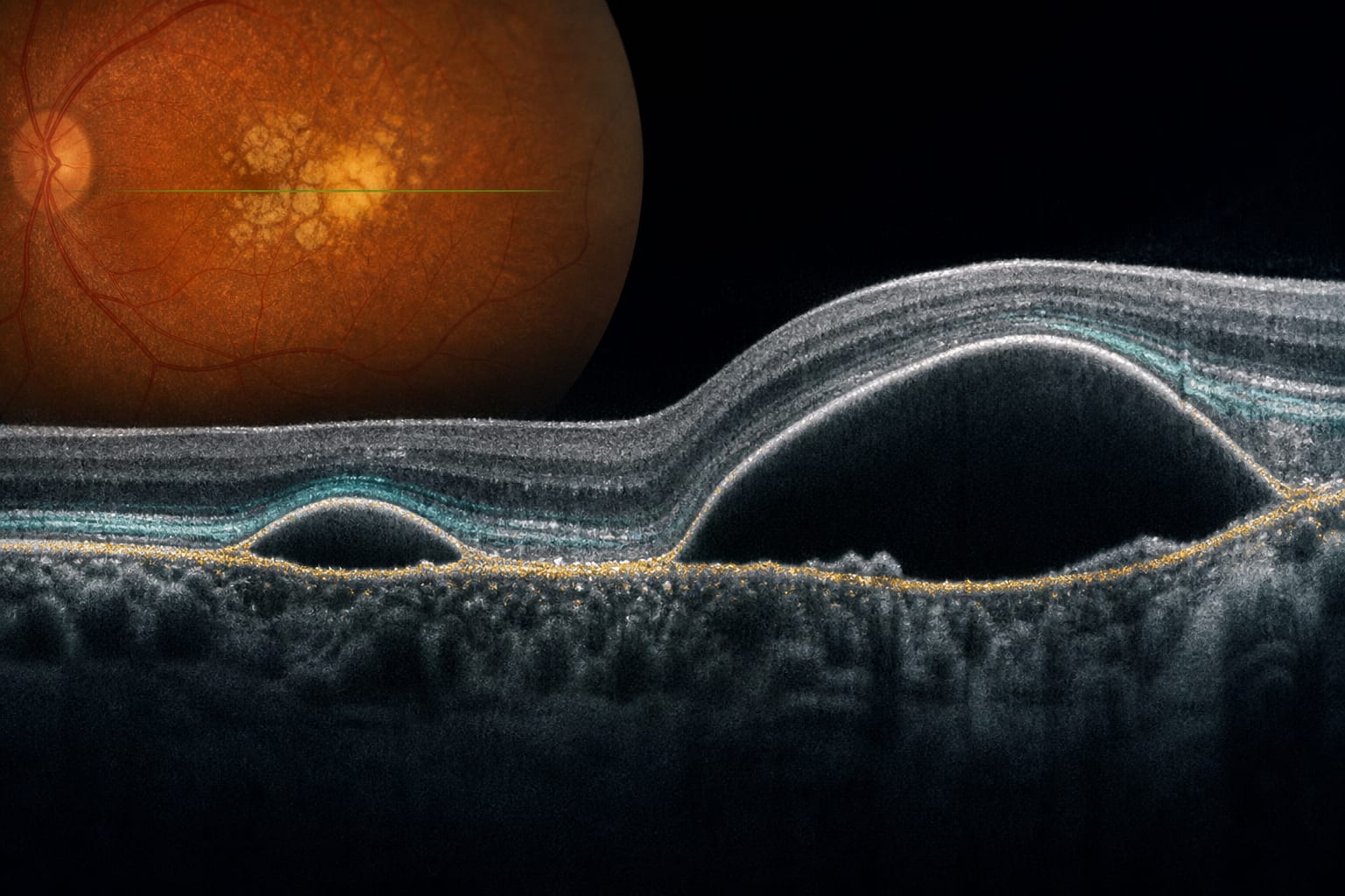
To describe a symptomatic free-floating vitreous cyst in a patient with longstanding floaters.
Approach:
Case: A 51-year-old woman had persistent left-eye floaters for more than 20 years, recently becoming more bothersome. Examination and multimodal imaging identified a mobile, pigmented cyst in the vitreous cavity.
Evaluation and treatment: The cyst was not attached to the optic disc or peripheral retina. There were no signs of intraocular inflammation, and serologic testing for parasitic infection was negative. Pars plana vitrectomy was performed.
Pathology and follow-up: The cyst wall was lined by pigmented cuboidal epithelial cells, supporting an origin from the iris or ciliary body pigmented epithelium. At 2 weeks, the patient reported symptom relief and visual acuity improved from 20/60 to 20/25.
Key Findings:
A free-floating vitreous cyst may be overlooked when persistent floaters are attributed to vitreous degeneration.
Histopathology supported an origin from the pigmented epithelium of the iris or ciliary body.
Symptoms improved after cyst removal in this case.
Interpretation:
The authors stated that vitreous cysts should be considered in the differential diagnosis of persistent floaters. They noted that vitrectomy may be considered in selected symptomatic cases, particularly when complete removal and histopathologic confirmation are desired.
Limitations:
The report describes a single patient.
Postoperative assessment was limited to 2 weeks, so the clinical course and surgical outcome should not be generalized beyond this case.
Conclusion:
The authors stated that a free-floating vitreous cyst should be considered in the differential diagnosis of persistent floaters.