Cross-software comparison shows strong agreement for quantitative indocyanine green fluorescence angiography in reconstructive surgery - Summary - MDSpire
Advertisement
Cross-software comparison shows strong agreement for quantitative indocyanine green fluorescence angiography in reconstructive surgery
To evaluate the agreement of key Q-ICG-FA parameters across two independent software platforms using identical ICG-FA recordings.
Approach:
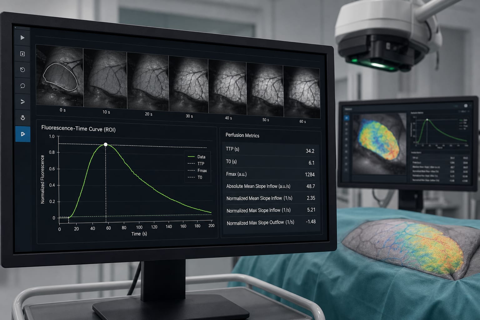
Study Design: Retrospective comparative analysis of 80 ICG-FA recordings from reconstructive procedures using two software platforms: AMS and EPA.
Data Analysis: Fluorescence-time curves (FTCs) were generated, and seven perfusion parameters were calculated. Agreement was assessed using intraclass correlation coefficients (ICC), non-parametric testing, and Bland–Altman analysis.
Key Findings:
Excellent agreement for time-to-peak (TTP) with ICC = 0.979 (95% CI: 0.967–0.987).
Normalized mean slope inflow showed good agreement with ICC = 0.944 (95% CI: 0.913–0.964).
Poor to moderate agreement for normalized maximum slopes with ICC values of 0.412 for inflow and 0.315 for outflow.
Significant systematic differences for six out of seven parameters, with AMS showing higher TTP values than EPA (p < 0.001).
Normalized mean slope inflow demonstrated the least variability and best agreement across platforms.
Interpretation:
TTP and normalized mean slope inflow may be reliable candidates for defining quantitative perfusion thresholds, although further clinical validation is needed.
Limitations:
Study limited to two software platforms and a single cohort of patients.
Potential variability in imaging protocols and analysis methods not fully addressed.
Conclusion:
Further clinical validation is necessary to establish reliable perfusion thresholds based on Q-ICG-FA parameters.
Acidic gum beat sugar-free at cranking out nitric oxide from beetroot juice — exactly backward from what test-tube studies predicted. Also this week: a sleep gene that ignores amyloid, and jackfruit sap moonlighting as a bone-building drug delivery system.