Conexiant’s news site is now MDSpire News. Learn more
Advertisement
Microinvasive Squamous Cell Carcinoma of the Cervix with Endometrial Involvement Misidentified as High-Grade Squamous Intraepithelial Lesion in a Postmenopausal Patient: A Case Study
To report a case of microinvasive cervical squamous cell carcinoma with endometrial spread that was misdiagnosed as high-grade squamous intraepithelial lesion (HSIL) during conization.
Approach:
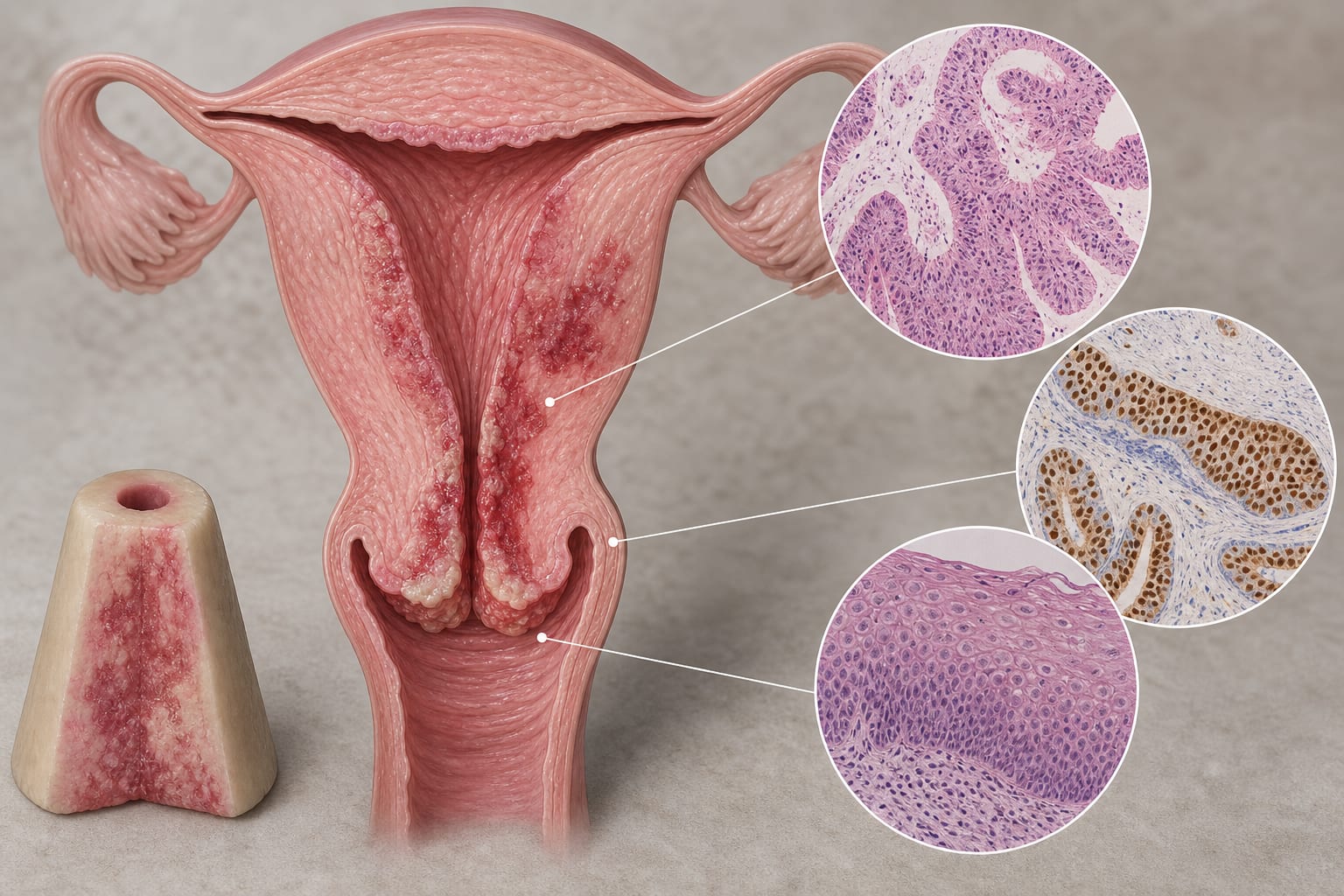
Case Presentation: A 56-year-old postmenopausal woman was referred after high-risk HPV positivity and cytology showing atypical squamous cells that could not rule out HSIL. Colposcopy-directed biopsy and subsequent deep cervical conization indicated HSIL/CIN grade 3 with glandular involvement, but no definite invasive carcinoma was identified. Due to concerns about residual disease, the patient underwent an extrafascial total hysterectomy.
Key Findings:
Final pathology revealed HSIL with a microinvasive squamous cell carcinoma focus measuring less than 0.5 mm and superficial endometrial spread.
Immunohistochemistry confirmed the cervical origin of the neoplasm, excluding primary endometrial disease.
At 6 months post-surgery, the patient was disease-free and tested negative for HPV.
Interpretation:
Conization may underestimate invasive disease in postmenopausal women, particularly with cervical atrophy and glandular involvement. Immunohistochemistry is crucial for distinguishing cervical neoplasia from primary endometrial lesions.
Limitations:
The findings are based on a single patient case, which limits the generalizability of the results.
The diagnostic challenges presented by cervical atrophy in postmenopausal women may not apply universally.
Conclusion:
This case highlights the importance of thorough evaluation of cervical lesions in postmenopausal women and the role of immunohistochemistry in accurate diagnosis.