To explore the potential of virtual biomarker staining as a scalable alternative to multiplex immunofluorescence (IF) for characterizing the tumor microenvironment.
Approach:
Key Findings:
Models demonstrated strong per-cell discrimination with AUCs ranging from 0.90 to 0.93.
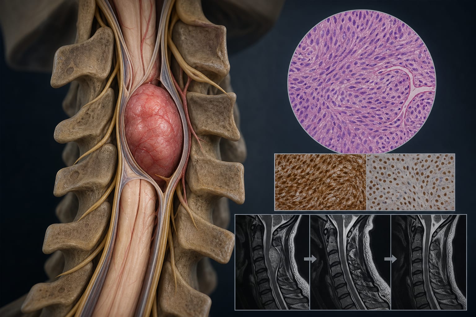
Virtual stains highlighted expected cell populations and tissue compartments as confirmed by pathologists.
Predictions maintained spatial context of biomarker expression.
Interpretation:
The findings support the feasibility of generating biologically meaningful virtual biomarker images from routine H&E whole-slide images, enhancing tumor microenvironment analysis.
Limitations:
Dependence on the quality of training data from clinical-grade IHC labels.
Potential variability in model performance across different tissue types.
Conclusion:
Virtual biomarker staining could revolutionize tumor microenvironment analysis in NSCLC by making it more practical, scalable, and accessible, potentially aiding in precision oncology workflows.