Ultrasound combined with serum parathyroid hormone for assessing autologous parathyroid graft viability after endoscopic thyroidectomy: a retrospective study - Summary - MDSpire
Advertisement
Ultrasound combined with serum parathyroid hormone for assessing autologous parathyroid graft viability after endoscopic thyroidectomy: a retrospective study
To investigate the diagnostic value of ultrasonography combined with serum parathyroid hormone (PTH) measurement in assessing the viability of autologous parathyroid grafts following endoscopic radical thyroidectomy for thyroid carcinoma.
Approach:
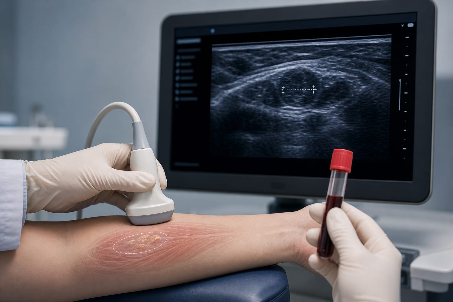
Study Design: A retrospective analysis of clinical data from 38 patients who underwent endoscopic unilateral radical thyroidectomy with autologous parathyroid transplantation.
Surgical Method: Parathyroid tissue was transplanted into the brachioradialis muscle using the homogenate injection method during surgery.
Assessment Methods: Postoperative PTH concentration ratios and ultrasonographic images of the graft site were evaluated to assess graft viability.
Key Findings:
PTH concentration on the transplantation side increased significantly post-surgery, peaking at 3 months with a 28.89-fold elevation compared to systemic circulation.
Ultrasonography at 3 months showed hypoechoic nodules at the graft site with blood flow signals detected by color Doppler flow imaging.
Interpretation:
The combination of ultrasonography and serum PTH concentration ratio provides evidence for parathyroid graft viability following autologous transplantation.
Limitations:
The study is limited to a retrospective analysis with a small sample size of 38 patients.
Findings may not be generalizable beyond the specific surgical technique and patient population studied.
Conclusion:
The study establishes a diagnostic approach for assessing graft viability using ultrasonography and serum PTH measurements.