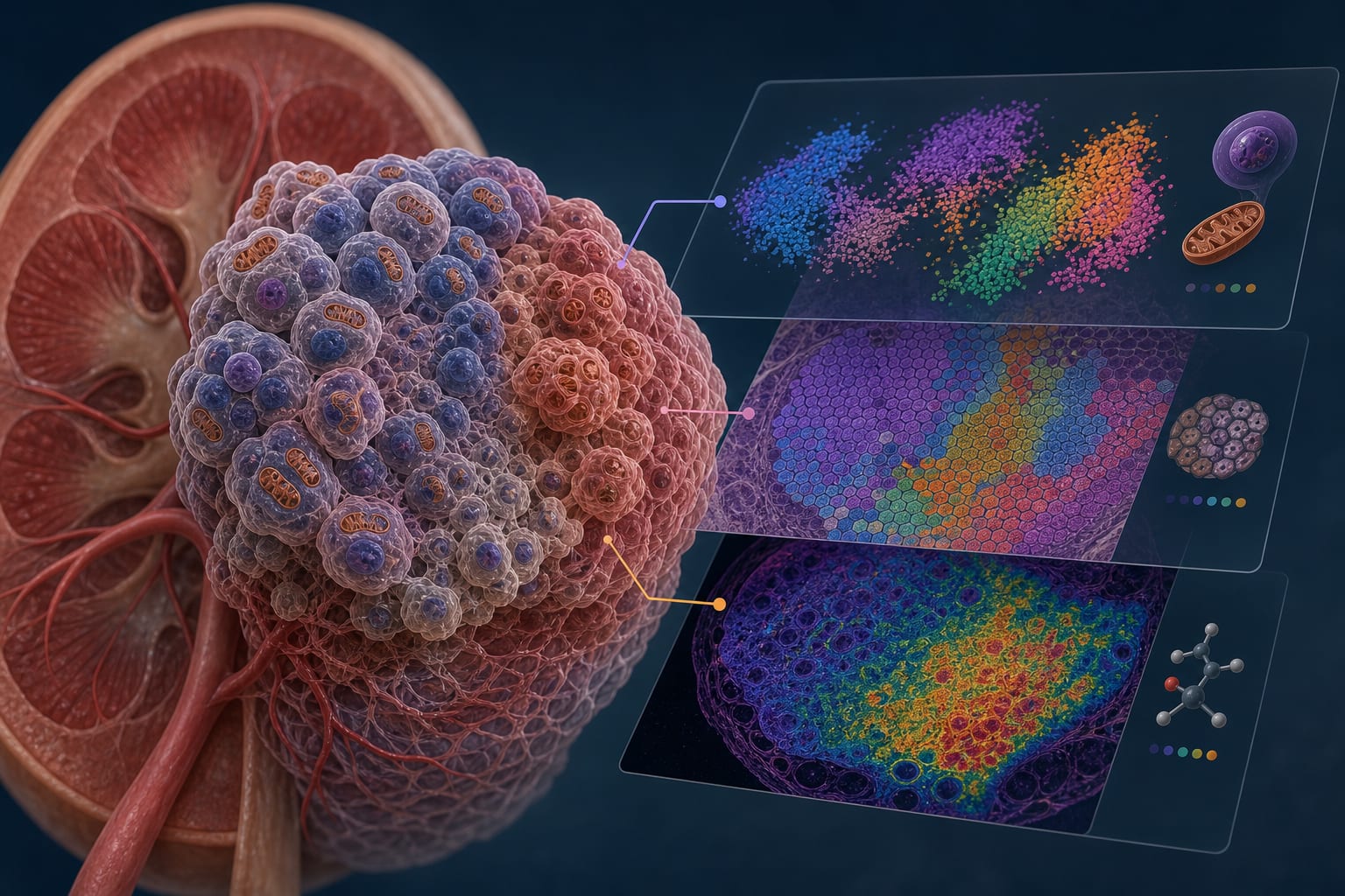
To determine whether radiomic features extracted from the peritumoral zone capture biologically relevant invasion-associated imaging phenotypes, using membrane-bound Hsp70 (mHsp70) as a molecular correlate.
Approach:
Key Findings:
The SVM classifier achieved an AUC of 0.875 in the independent test cohort.
Higher-order texture features were identified as major contributors to model predictions.
The radiomics-derived Invasion Burden Index (IBI) showed a significant positive correlation with mHsp70 expression (Spearman ρ = 0.67, p = 0.0004).
Regions with elevated invasion-associated radiomic signatures often extended beyond conventional MRI-defined tumor margins.
Interpretation:
Voxel-wise radiomic analysis identifies invasion-associated imaging phenotypes that correlate with membrane Hsp70 expression, a biological marker associated with aggressive tumor behavior.
Limitations:
The study is retrospective and may be subject to biases inherent in such designs.
Further prospective studies with spatially matched biological validation are needed.
Conclusion:
Radiomic invasion mapping may serve as a non-invasive tool for characterizing infiltrative glioma biology.
by Natalia Mikhailova, Anastasiia Nechaeva, Danila Bobkov, Hem Chandra Jha, Kadambari KV, Yerramneni Vamsi Krishna, Hari Ponnamma Rani, Aleksander Kim, Maxim Shevtsov