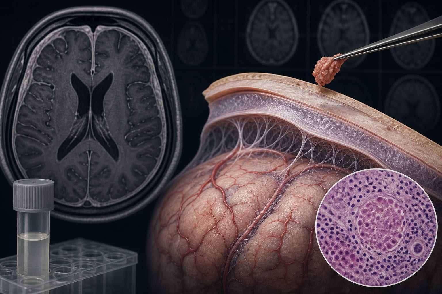
To describe a diagnostically challenging case of paucibacillary tuberculous meningitis (TBM) characterized by atypical presentation, negative microbiological testing, and clinical-radiological dissociation.
Approach:
Case Presentation: A 63-year-old woman with newly diagnosed diabetes presented with sudden headache, dysarthria, right-sided weakness, and focal seizures, without fever, meningeal signs, or respiratory symptoms. Initial imaging and near-normal cerebrospinal fluid findings suggested viral meningoencephalitis.
Diagnostic Process: CSF GeneXpert MTB/RIF, mycobacterial culture, and metagenomic next-generation sequencing were negative, and the Marais score was below the threshold for possible TBM. After clinical symptoms resolved but MRI abnormalities progressed, meningeal biopsy showed granulomatous inflammation with a single acid-fast bacillus, and peripheral-blood interferon-gamma release assay was positive, strongly supporting TBM.
Treatment and Follow-up: Anti-tuberculosis therapy was given for approximately 12 months, with dexamethasone administered for more than 3 months. Follow-up MRI at 9 months showed almost complete resolution of meningeal enhancement and cerebral edema, and the patient achieved full neurological recovery.
Key Findings:
Paucibacillary TBM can occur with normal or near-normal CSF findings and negative molecular and microbiological tests.
The patient’s Marais diagnostic score was 2, below the score of 6 required for possible TBM under those criteria.
Meningeal biopsy was pivotal in establishing a highly probable diagnosis when CSF molecular testing remained negative.
Interpretation:
This case illustrates that TBM can mimic stroke, viral encephalitis, or noninfectious conditions and that symptomatic improvement does not necessarily indicate disease control. Radiological progression despite clinical quiescence should prompt strong consideration of meningeal biopsy, particularly when molecular tests on CSF remain negative.
Limitations:
Near-normal CSF findings contributed to delayed suspicion of TBM.
The lack of paired serum glucose precluded accurate calculation of the CSF-to-serum glucose ratio.
GeneXpert MTB/RIF, mycobacterial culture, and metagenomic next-generation sequencing of CSF were all negative despite the eventual diagnosis.
HSV/VZV infection, neurosarcoidosis, and nontuberculous mycobacterial infection were difficult to exclude before treatment but were considered less likely after substantial radiological improvement with anti-tuberculosis therapy and sustained remission off treatment.
Conclusion:
Paucibacillary TBM may present without typical infectious features and remain undetected by contemporary CSF microbiological testing. Complete symptomatic improvement does not exclude TBM, and radiological progression despite clinical quiescence should prompt strong consideration of meningeal biopsy, particularly when molecular tests on CSF remain negative.
Investigators compared intra-articular microbial profiles and predicted metabolic pathways in patients with hip osteoarthritis and nonarthritic controls.