To identify clinical and high-resolution vessel wall magnetic resonance imaging features associated with ischemic stroke and to develop a patient-level model for short-term risk prediction.
Approach:
Statistical Analysis: Applied LASSO regression for variable selection and mixed-effects logistic regression to identify factors associated with ischemic events, ensuring robust statistical analysis.
Key Findings:
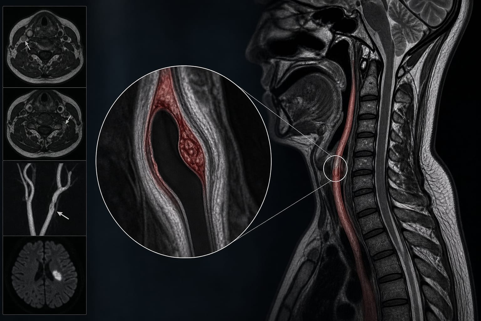
At the vessel level, white blood cell count, intraluminal thrombus, severe stenosis or occlusion, and alcohol consumption were independently associated with ischemic events.
At the patient level, WBC count, intraluminal thrombus, male sex, and alcohol consumption were independent predictors of ischemic stroke.
The nomogram demonstrated good discriminative ability with an optimism-corrected area under the curve of 0.837 (95% CI: 0.810–0.852).
Interpretation:
The patient-level model shows good performance in predicting ischemic stroke risk in patients with CeAD.
Limitations:
Retrospective design may introduce selection bias.
Findings may not be generalizable to all populations due to the specific patient cohort.
Conclusion:
The developed nomogram may assist in early risk stratification and individualized clinical decision-making for patients with CeAD.