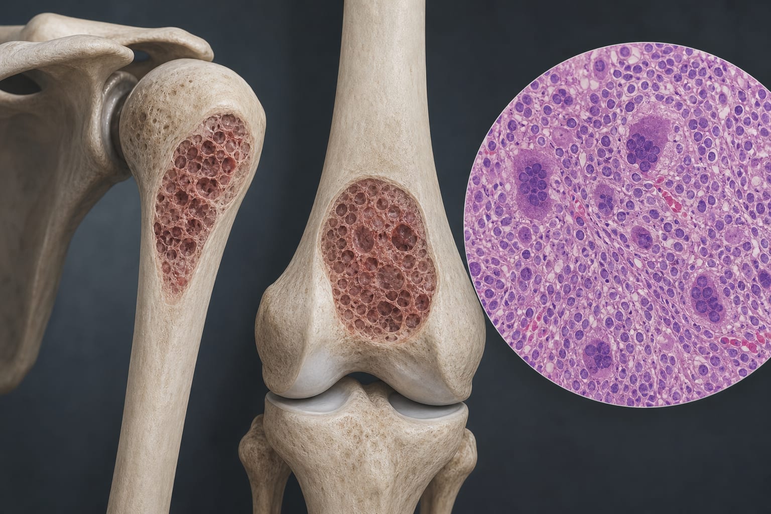
To report two rare cases of giant cell reparative granuloma (GCRG) in the proximal humerus and distal femur, highlighting their atypical skeletal manifestations and diagnostic challenges.
Approach:
Case Report 1: A 45-year-old woman with left shoulder pain and a history of heavy lifting underwent imaging that revealed a well-defined osteolytic lesion in the proximal humerus. Surgical intervention confirmed GCRG through histopathological examination.
Case Report 2: A 26-year-old man presented with knee pain after a fall. Imaging showed an osteolytic lesion in the distal femur, initially suspected to be a giant cell tumor. Surgical intervention confirmed GCRG.
Key Findings:
GCRG is a rare benign lesion that typically arises in the mandible and maxilla, with extremity involvement being extremely uncommon.
Both cases presented with a history of prior trauma and imaging findings that mimicked giant cell tumors of bone.
Histopathological examination post-surgery confirmed the diagnosis of GCRG in both cases.
Interpretation:
These cases highlight the importance of considering GCRG in the differential diagnosis of osteolytic lesions at atypical sites, particularly in patients with a history of trauma.
Limitations:
The rarity of GCRG in long bones may limit generalizability of findings.
The cases are based on a small sample size, which may not represent broader trends.
Conclusion:
Accurate diagnosis of GCRG requires integration of radiologic and histopathological findings to avoid misdiagnosis.