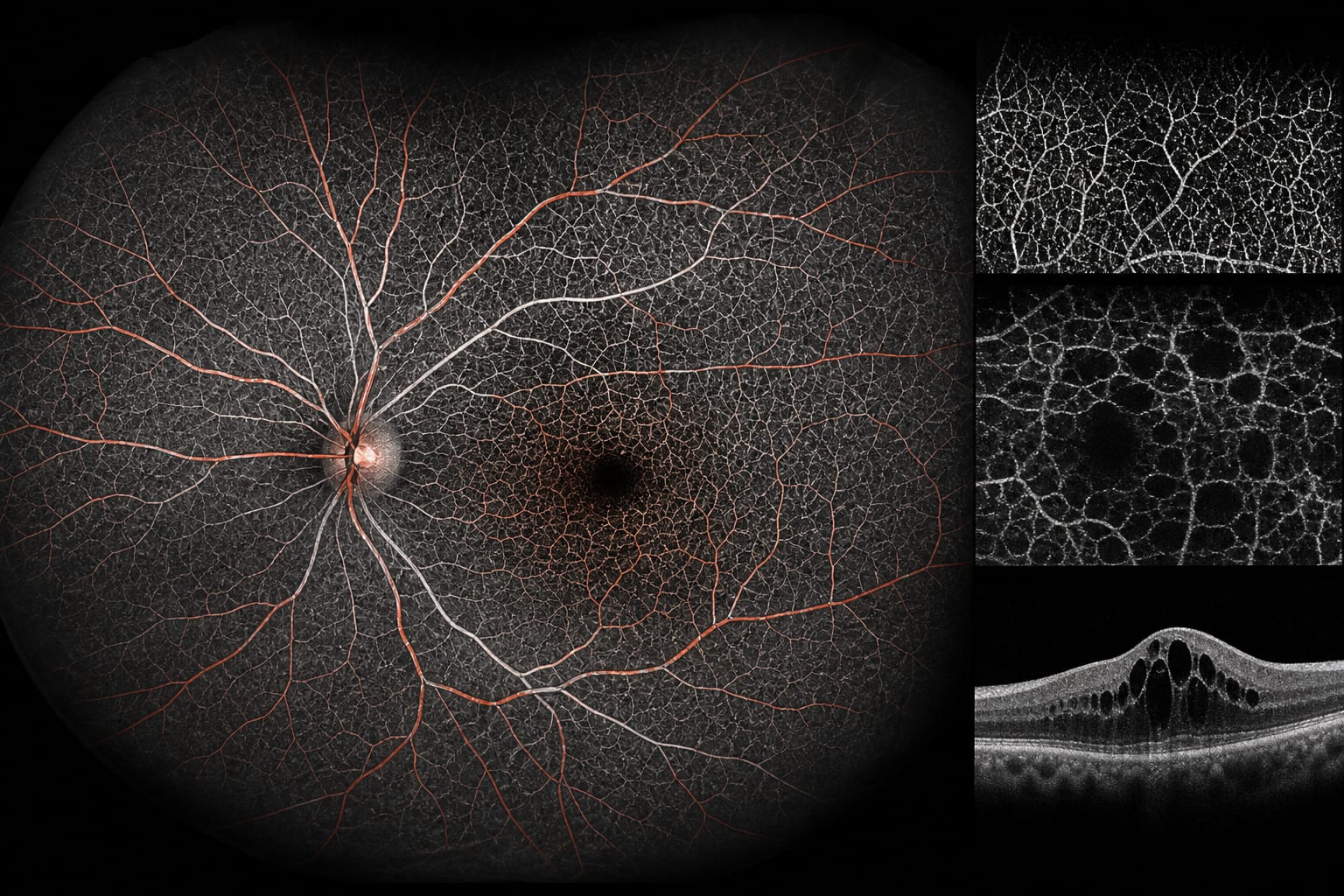
To investigate retinal blood flow alterations in diabetic macular edema (DME) using ultra-widefield optical coherence tomography angiography (OCTA), highlighting its significance in early detection.
Key Findings:
Nasal SVC vascular density was significantly reduced in diabetes patients without retinopathy (p<0.05).
NPDR patients exhibited widespread SVC vascular density reduction compared to controls and DM patients (p<0.01).
DME patients showed increased SVC vascular density in the peripheral retina (p<0.05).
DVC changes were significant only in the macular area in NPDR patients (p<0.05).
Peripheral DVC vascular density strongly correlated with DME presence (r=0.85, p<0.01).
Interpretation:
Nasal SVC vascular alterations are detectable early in preclinical diabetic retinopathy, and peripheral DVC vascular density correlates closely with DME, suggesting potential for early intervention.
Limitations:
The study is retrospective and may be subject to selection bias, including potential confounding factors.
Limited sample size may affect the generalizability of the findings.
Conclusion:
The study highlights the potential of OCTA in detecting early vascular changes associated with diabetic macular edema, paving the way for future research on therapeutic interventions.
US claims data showed rising prevalence of diabetic retinal disease in type 1 and type 2 diabetes, while incidence declined in type 1 diabetes and moved closer to type 2 rates by 2022.