To compare Microvascular Flow imaging (MV-Flow) with Color Doppler Flow Imaging (CDFI) and evaluate a machine learning (ML) framework integrating MV-Flow parameters for differentiating breast lesions, highlighting the importance of accurate diagnosis in breast cancer management.
Approach:
Key Findings:
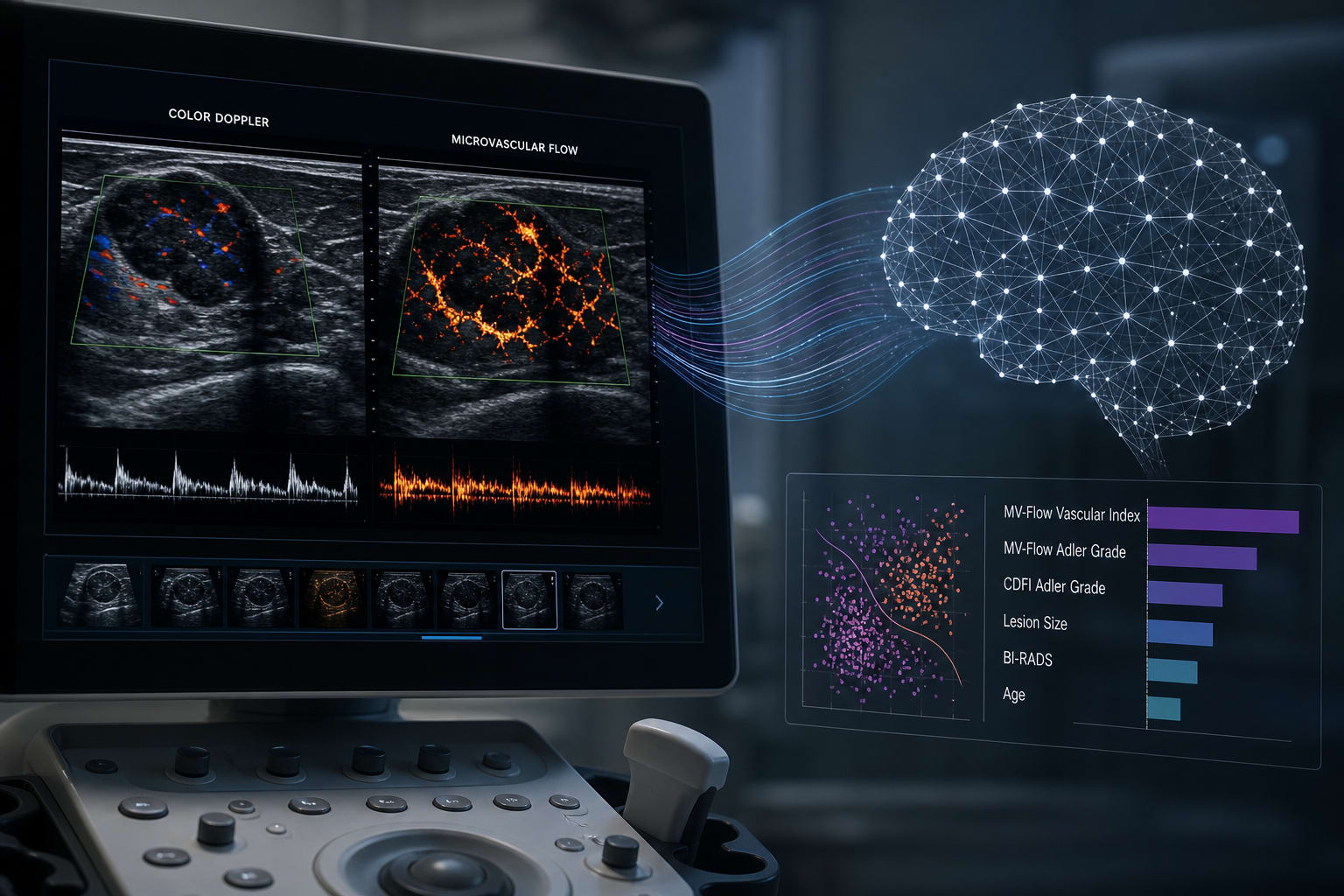
MV-Flow detected blood flow in 16 lesions missed by CDFI, indicating its superior sensitivity.
Higher inter-observer agreement for MV-Flow (weighted Kappa=0.68) compared to CDFI (0.51), suggesting improved reliability.
Median Vascular Index (VI) was significantly higher in malignant lesions (20.25) than benign ones (3.10, P<0.001), emphasizing its diagnostic value.
Diagnostic AUC for MV-Flow Adler grade, VI alone, and their combination were 0.874, 0.823, and 0.888, respectively, indicating strong diagnostic performance.
K-Nearest Neighbors model achieved the best performance with an accuracy of 0.927 and an F1-score of 0.947, showcasing the effectiveness of the ML approach.
Interpretation:
SHAP analysis identified BI-RADS category and patient age as the most important predictive features in the ML model, which could guide future diagnostic strategies.
Limitations:
Potential biases in patient selection and the generalizability of findings should be acknowledged.
Conclusion:
MV-Flow outperforms CDFI in depicting breast tumor microvasculature, and ML models integrating MV-Flow parameters can optimize diagnostic accuracy.